Coloured SEM of the aortic valve
Numéro d’image : 11868998
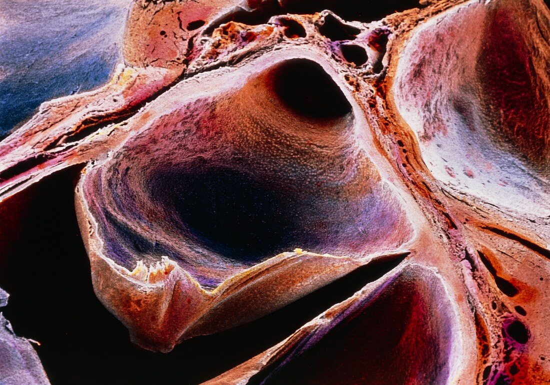
| Coloured scanning electron micrograph of the aortic valve. The heart was sectioned at the level of the coronary sulcus,which separates the atria from the ventricles,and the valve is seen from above. The aortic valve is found between the left ventricle and the aorta and prevents the flow of blood back into the left ventricle. It is formed by three semilunar cusps; only two are visible here,one is the large area at centre and the other one is at bottom right. The dark round area at top centre is the orifice of the left coronary artery; a semilunar cusp of the pulmonary valve is at top right. Magnification: x25 at 6x7cm size | |
| Licence : | Droits gérés |
| Crédit: | Science Photo Library / PROFESSORS P.M. MOTTA & G. MACCHIARELLI |
| Taille de l’image : | 5030 px × 3525 px |
| Model Release : | Non requis |
| Property Release : | Non requis |
| Restrictions : | - |
Prix pour cette image À partir de 45 €
Produit vendu
(Calendrier, Carte postale, Carte de vœux, Impression sur textile, Packaging etc)
À partir de 45 €
Usage commercial
(Affichage, Annonce presse, Annonce TV, Carte, Digital - hors rés. sociaux, Digital - rés. sociaux etc)
À partir de 45 €
Éditorial
(Digital, Journal, Livre, Livre pratique, Magazine, Télévision etc)
À partir de 60 €
Usage non-commercial
(Digital - hors rés. sociaux, Digital - rés. sociaux etc)
À partir de 120 €