F/colour CT scan of chest showing heart and lungs
Numéro d’image : 11868989
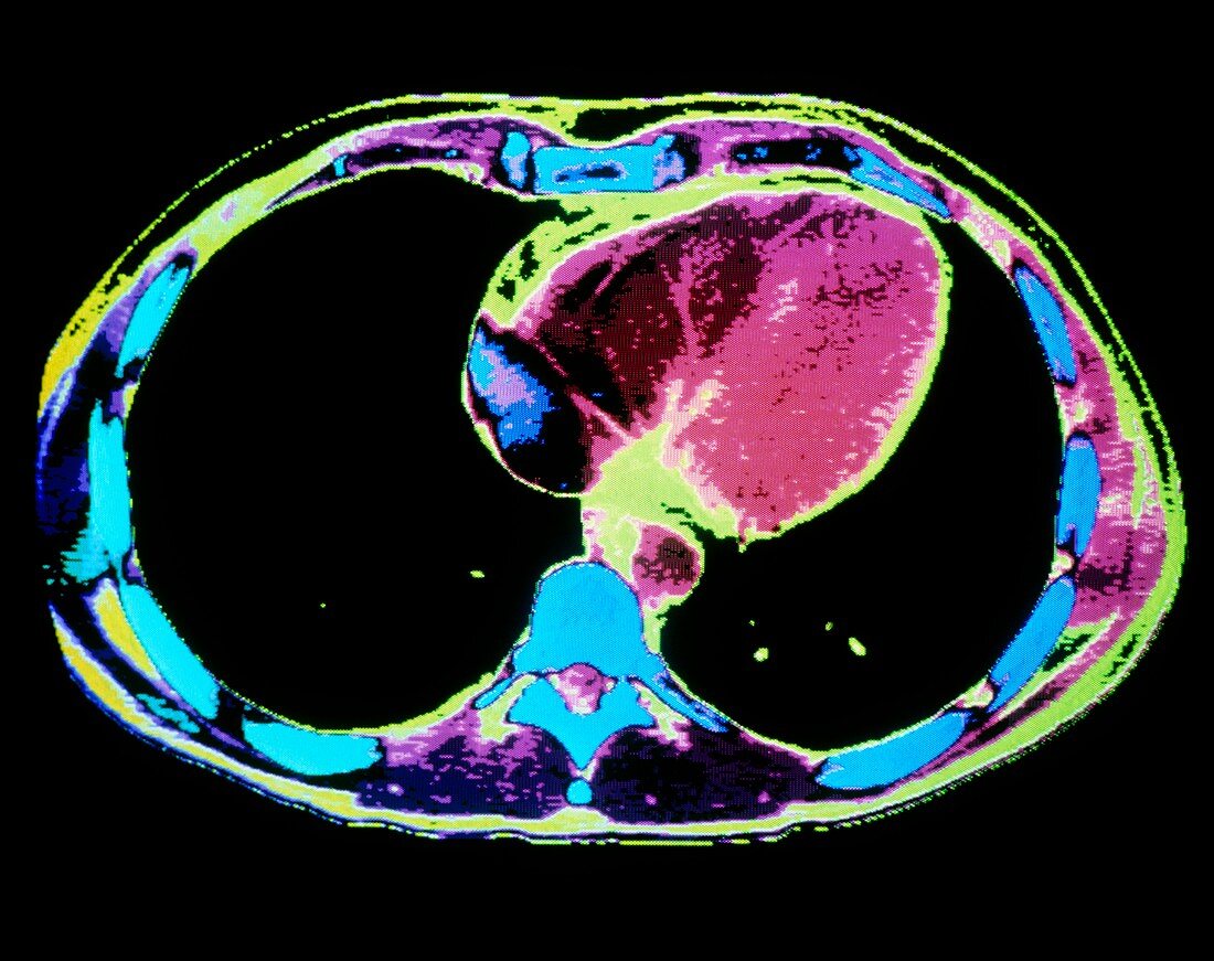
| Heart and lungs. False-colour computed tomography (CT) scan of an axial section through the chest,showing a healthy heart. At centre right is the heart (pink); the descending aorta (lower centre,circular pink) is a major artery from the heart that supplies blood to the body. On either side are the lung fields (coloured black). A cross- section of a thoracic vertebra of the spine (blue) appears below the aorta; circling the lungs are rib bones (blue). CT scans are obtained by taking a series of X-ray images from points on a circle around the subject. A computer is used to compare and arrange the resulting data into an image "slice" through the body | |
| Licence : | Droits gérés |
| Crédit: | Science Photo Library / GCA |
| Taille de l’image : | 4776 px × 3780 px |
| Model Release : | Non requis |
| Property Release : | Non requis |
| Restrictions : | - |
Prix pour cette image À partir de 45 €
Produit vendu
(Calendrier, Carte postale, Carte de vœux, Impression sur textile, Packaging etc)
À partir de 45 €
Usage commercial
(Affichage, Annonce presse, Annonce TV, Carte, Digital - hors rés. sociaux, Digital - rés. sociaux etc)
À partir de 45 €
Éditorial
(Digital, Journal, Livre, Livre pratique, Magazine, Télévision etc)
À partir de 60 €
Usage non-commercial
(Digital - hors rés. sociaux, Digital - rés. sociaux etc)
À partir de 120 €