False-colour SEM of a portion of a cardiac valve
Numéro d’image : 11868987
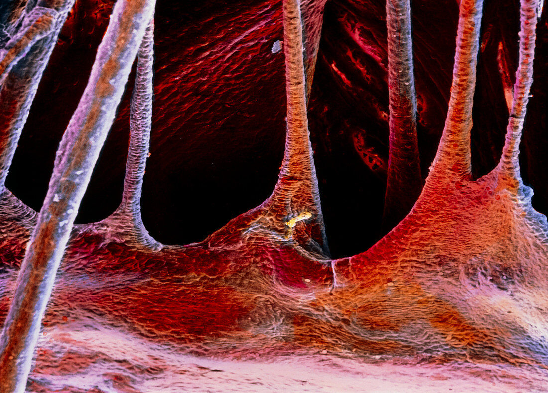
| Heart valve. False-colour scanning electron micrograph of a portion of one of the atrioventricular valves of the heart: either the tricuspid or the mitral valve. They channel the flow of blood from the atria to the ventricles and prevent any backflow. The image clearly shows the chordae tendineae,the fibrous filiform collagenous cords,which connect the cusps of the valve to the papillary muscles of the heart. The epithelium at top is known as the endocardium: it is formed by a single layer of flattened cells supported by fibro-elastic connective tissue. Magnification: x70 at 6x7cm size. x115 at 4x5ins | |
| Licence : | Droits gérés |
| Crédit: | Science Photo Library / UNIVERSITY LA SAPIENZA, ROME / DEPT. OF ANATOMY / PROF. P. MOTTA |
| Taille de l’image : | 5197 px × 3734 px |
| Model Release : | Non requis |
| Property Release : | Non requis |
| Restrictions : | - |
Prix pour cette image À partir de 45 €
Produit vendu
(Calendrier, Carte postale, Carte de vœux, Impression sur textile, Packaging etc)
À partir de 45 €
Usage commercial
(Affichage, Annonce presse, Annonce TV, Carte, Digital - hors rés. sociaux, Digital - rés. sociaux etc)
À partir de 45 €
Éditorial
(Digital, Journal, Livre, Livre pratique, Magazine, Télévision etc)
À partir de 60 €
Usage non-commercial
(Digital - hors rés. sociaux, Digital - rés. sociaux etc)
À partir de 120 €