NMR scan of a normal chest & heart
Numéro d’image : 11868958
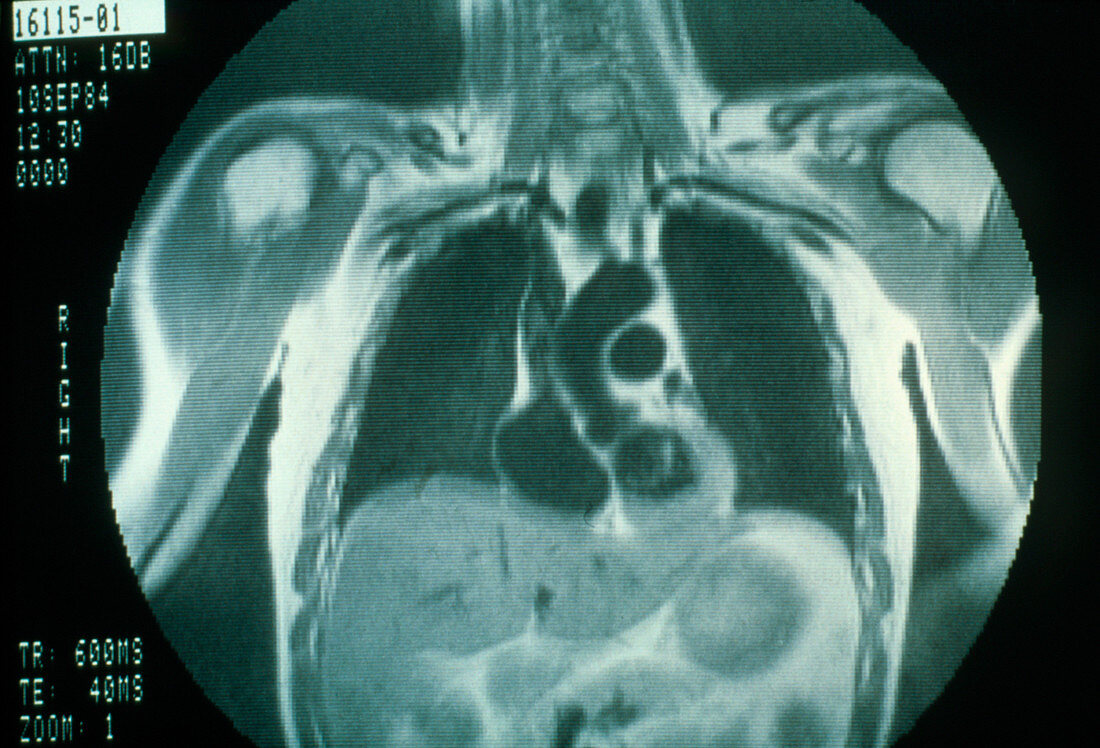
| Nuclear magnetic resonance (NMR) image of the chest area of a normal healthy subject,featuring the heart at the end of systole; the heart appears at centre right,to the left of the lower lobe of the right lung. Systole is the period of the cardiac cycle when the heart contracts (the term normally refers to ventricular systole,which lasts about 0.3 seconds); the blood-filled ventricles are pumping blood to the lungs and around the general circulation. Also visible are both lungs,the ribs (at far edges of both lungs),the carotid and subclavian arteries (blood vessels supplying the head and arms respectively) and vertebrae of the neck | |
| Licence : | Droits gérés |
| Crédit: | Science Photo Library |
| Taille de l’image : | 3648 px × 2480 px |
| Model Release : | Non requis |
| Property Release : | Non requis |
| Restrictions : | - |
Prix pour cette image À partir de 45 €
Produit vendu
(Calendrier, Carte postale, Carte de vœux, Impression sur textile, Packaging etc)
À partir de 45 €
Usage commercial
(Affichage, Annonce presse, Annonce TV, Carte, Digital - hors rés. sociaux, Digital - rés. sociaux etc)
À partir de 45 €
Éditorial
(Digital, Journal, Livre, Livre pratique, Magazine, Télévision etc)
À partir de 60 €
Usage non-commercial
(Digital - hors rés. sociaux, Digital - rés. sociaux etc)
À partir de 120 €