SEM of capillaries of the gall bladder
Numéro d’image : 11868920
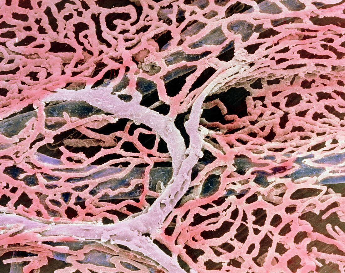
| Capillaries of the gall bladder. Coloured Scanning Electron Micrograph (SEM) of blood vessels lining the wall of the gall bladder. Here,a rich network of fine capillaries is seen (pink) branching off a blood vessel (grey). Beneath this network are a number of larger blood vessels (blue). This vascular system occurs in the submucosal layer of the gall bladder. Blood and lymphatic vessels serve to drain water reabsorbed from bile during bile formation. The gall bladder is a muscular sac,attached to the liver,which collects and concentrates bile,a substance which aids in fat digestion. Magnification: x100 at 6x7cm size. Magnification: x125 at 4x5 inch size | |
| Licence : | Droits gérés |
| Crédit: | Science Photo Library / PROFESSOR P.M. MOTTA, A. CAGGIATI & G. MACCHIARELLI |
| Taille de l’image : | 3661 px × 2899 px |
| Model Release : | Non requis |
| Property Release : | Non requis |
| Restrictions : | - |
Prix pour cette image À partir de 45 €
Produit vendu
(Calendrier, Carte postale, Carte de vœux, Impression sur textile, Packaging etc)
À partir de 45 €
Usage commercial
(Affichage, Annonce presse, Annonce TV, Carte, Digital - hors rés. sociaux, Digital - rés. sociaux etc)
À partir de 45 €
Éditorial
(Digital, Journal, Livre, Livre pratique, Magazine, Télévision etc)
À partir de 60 €
Usage non-commercial
(Digital - hors rés. sociaux, Digital - rés. sociaux etc)
À partir de 120 €