Blood vessels in the intestine,SEM
Numéro d’image : 11868807
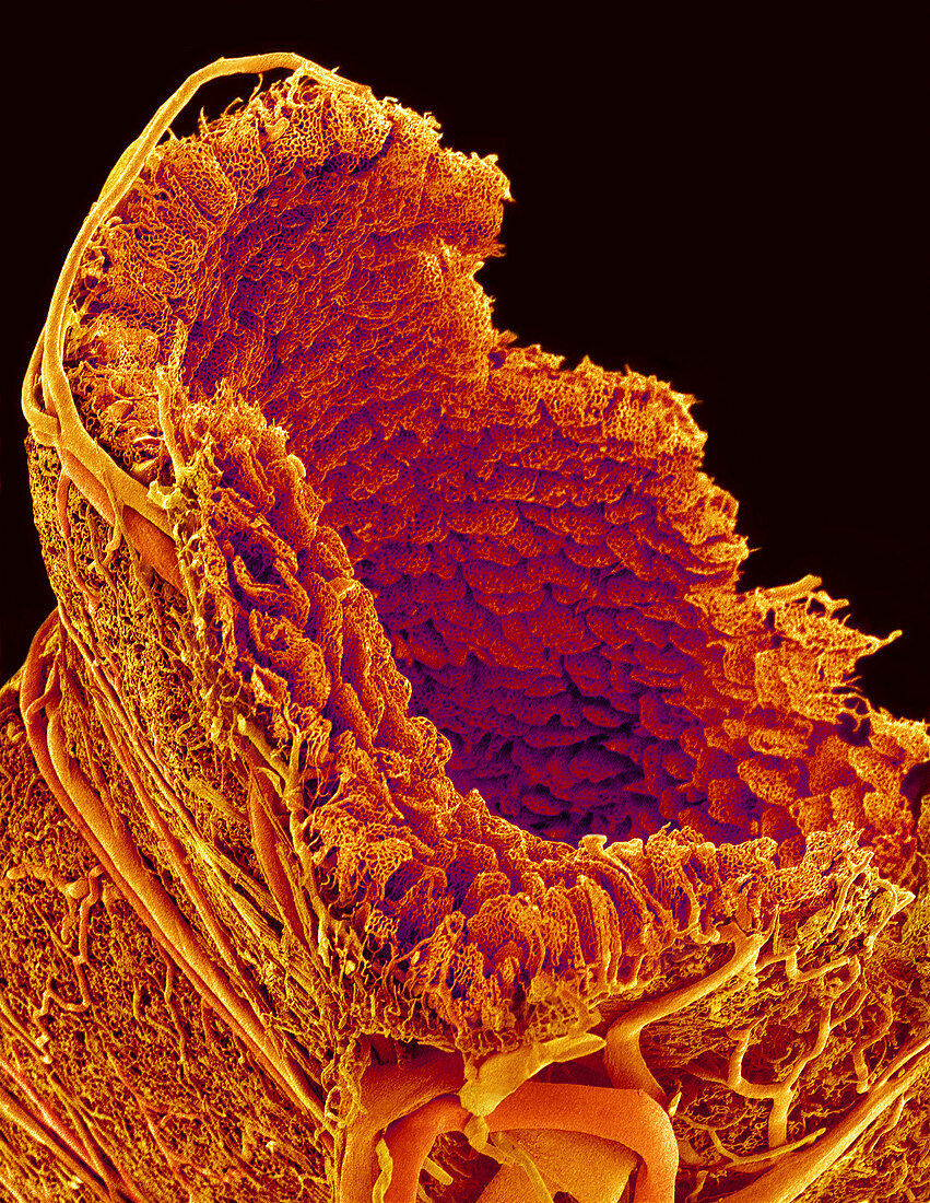
| Blood vessels of the small intestine,coloured scanning electron micrograph (SEM). Large blood vessels (bottom) supply the intestine and encircle its outer wall (lower frame and upper left). The inner wall of the intestine is folded into villi,finger-like projections that increase the surface area of the inner wall,aiding the absorption of nutrients from food. The villi contain a network of thin capillaries,through which nutrients pass into the blood. This is a cast: the vessels were filled with a resin,which then set. The surrounding tissues were then eaten away chemically,leaving the cast of the vessels. Magnification: x21 when printed 10cm high | |
| Licence : | Droits gérés |
| Crédit: | Science Photo Library / Nishinaga, Susumu |
| Taille de l’image : | 2894 px × 3741 px |
| Model Release : | Non requis |
| Property Release : | Non requis |
| Restrictions : | - |
Prix pour cette image À partir de 45 €
Produit vendu
(Calendrier, Carte postale, Carte de vœux, Impression sur textile, Packaging etc)
À partir de 45 €
Usage commercial
(Affichage, Annonce presse, Annonce TV, Carte, Digital - hors rés. sociaux, Digital - rés. sociaux etc)
À partir de 45 €
Éditorial
(Digital, Journal, Livre, Livre pratique, Magazine, Télévision etc)
À partir de 60 €
Usage non-commercial
(Digital - hors rés. sociaux, Digital - rés. sociaux etc)
À partir de 120 €
Mots clés
- anatomie,
- anatomique,
- approvisionnement,
- biologie,
- biologique,
- brun,
- capillaires,
- circulatoire,
- coloré,
- colorié,
- colorisé,
- corps humain,
- duodénal,
- duodénum,
- intestin grêle,
- intestinal,
- jeter,
- lit capillaire,
- M.E.B.,
- MEB,
- microscope électronique à balayage,
- résine,
- sang,
- système,
- système digestif,
- vaisseau,
- vaisseau sanguin,
- vaisseaux,
- vasculaire,
- villosité,
- villosités