Coloured SEM of section through a human arteriole
Numéro d’image : 11868601
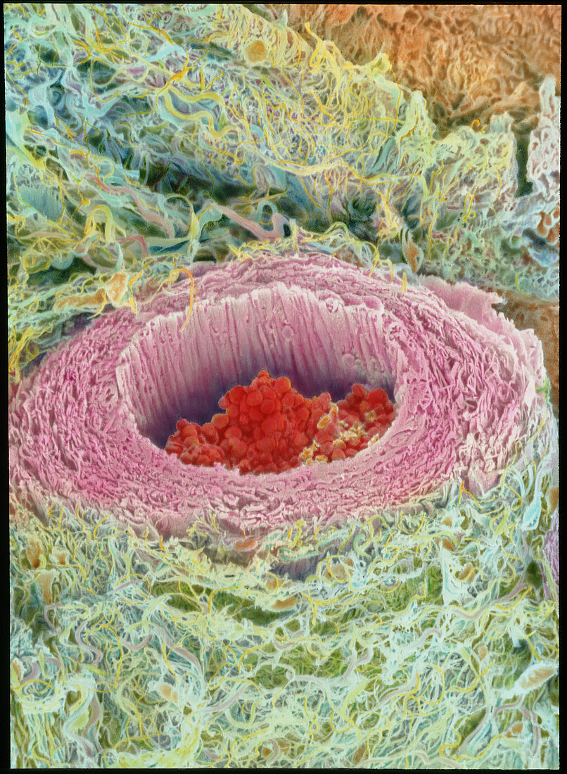
| Arteriole. Coloured scanning electron micrograph (SEM) of a cross-section through a small human artery known as an arteriole. This blood vessel reveals red blood cells travelling through its central lumen. Its inner wall (tunica intima) is thin and appears folded,comprising an endothelial lining and a thin elastic layer which stretches. A thicker tunica media layer (pink) makes up much of the wall of the arteriole,and is almost entirely composed of smooth muscle cells. An outer tunica adventitia layer merges with surrounding connec- tive tissue (blue). These layers enable the arteriole to expand and contract,regulating the arterial blood pressure. Magnification: unknown | |
| Licence : | Droits gérés |
| Crédit: | Science Photo Library / Gschmeissner, Steve |
| Taille de l’image : | 3201 px × 4369 px |
| Model Release : | Non requis |
| Property Release : | Non requis |
| Restrictions : | - |
Prix pour cette image À partir de 45 €
Produit vendu
(Calendrier, Carte postale, Carte de vœux, Impression sur textile, Packaging etc)
À partir de 45 €
Usage commercial
(Affichage, Annonce presse, Annonce TV, Carte, Digital - hors rés. sociaux, Digital - rés. sociaux etc)
À partir de 45 €
Éditorial
(Digital, Journal, Livre, Livre pratique, Magazine, Télévision etc)
À partir de 60 €
Usage non-commercial
(Digital - hors rés. sociaux, Digital - rés. sociaux etc)
À partir de 120 €
Mots clés
- agrandissement,
- anatomie,
- artère,
- artériole,
- biologie humaine,
- cellule sanguine,
- corps humain,
- globule rouge,
- histologie,
- images,
- intima,
- M.E.B.,
- MEB,
- media,
- microscope électronique à balayage,
- petit,
- photos au microscope,
- sang,
- sujets,
- tunica intima,
- tunica media,
- tunique intermédiaire d'un vaisseau artériel,
- tunique interne d'un vaisseau sanguin,
- vaisseau,
- vaisseau sanguin,
- vaisseaux