Coloured SEM of blood vessels in human oviduct
Numéro d’image : 11868582
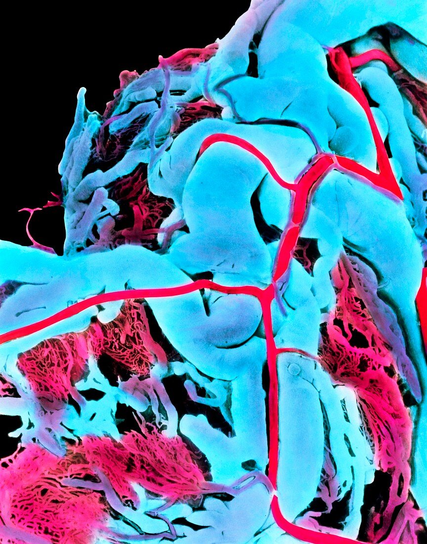
| Blood vessels of human oviduct. Coloured Scanning Electron Micrograph (SEM) of a vascular corrosion cast of blood vessels on the surface of the human oviduct. Tissue around the blood vessels has been removed. Large external veins (blue) are visible,with branches of the tubal artery (pink) seen. A bed of fine capillaries is positioned beneath the larger blood vessels. The oviduct or Fallopian tube is the channel which conducts the female egg from the ovary to the womb,and in which the egg is usually fertilized. Here,the oviduct is highly vascularised 24 hours after an egg was released at ovulation. Magnification: x15 at 6x7cm size. x50 at 8x10ins | |
| Licence : | Droits gérés |
| Crédit: | Science Photo Library / PROFESSOR P.M. MOTTA & E. VIZZA |
| Taille de l’image : | 3757 px × 4783 px |
| Model Release : | Non requis |
| Property Release : | Non requis |
| Restrictions : | - |
Prix pour cette image À partir de 45 €
Produit vendu
(Calendrier, Carte postale, Carte de vœux, Impression sur textile, Packaging etc)
À partir de 45 €
Usage commercial
(Affichage, Annonce presse, Annonce TV, Carte, Digital - hors rés. sociaux, Digital - rés. sociaux etc)
À partir de 45 €
Éditorial
(Digital, Journal, Livre, Livre pratique, Magazine, Télévision etc)
À partir de 60 €
Usage non-commercial
(Digital - hors rés. sociaux, Digital - rés. sociaux etc)
À partir de 120 €