Coloured SEM of an artery in tongue tissue
Numéro d’image : 11868581
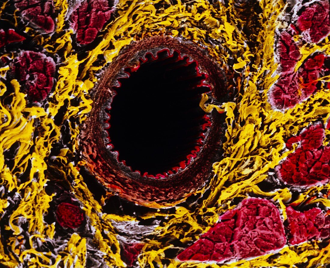
| Artery. Coloured Scanning Electron Micrograph (SEM) of a cross-section through an artery. At centre is the dark lumen of the artery through which blood passes. Its inner wall (tunica intima) is folded due to contraction of the artery. The artery has two outer layers: tunica media (dark brown) composed of smooth muscle cells; tunica adventitia (light brown) which is elastic. Around the artery is connective tissue (yellow),and bands of striated muscle fibres (red). This striated muscle tissue was found in the tongue. Magnification: x170 at 6x7cm size. Magnification: x220 at 4x5 inch size | |
| Licence : | Droits gérés |
| Crédit: | Science Photo Library / UNIVERSITY LA SAPIENZA, ROME / DEPT. OF ANATOMY / PROF. P. MOTTA |
| Taille de l’image : | 4692 px × 3803 px |
| Model Release : | Non requis |
| Property Release : | Non requis |
| Restrictions : | - |
Prix pour cette image À partir de 45 €
Produit vendu
(Calendrier, Carte postale, Carte de vœux, Impression sur textile, Packaging etc)
À partir de 45 €
Usage commercial
(Affichage, Annonce presse, Annonce TV, Carte, Digital - hors rés. sociaux, Digital - rés. sociaux etc)
À partir de 45 €
Éditorial
(Digital, Journal, Livre, Livre pratique, Magazine, Télévision etc)
À partir de 60 €
Usage non-commercial
(Digital - hors rés. sociaux, Digital - rés. sociaux etc)
À partir de 120 €