LM of normal human cartilage
Numéro d’image : 11868484
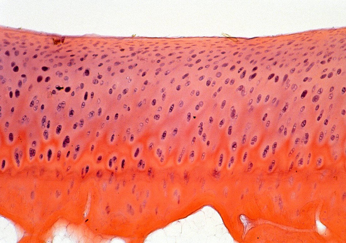
| Light micrograph of normal human cartilage at the surface of an articulated bone joint. Cartilage consists of cells,called chondrocytes,seen here as dark purple,which secrete a matrix of collagen fibres and acidic polysaccharides. The retention of a cartilage layer at the surface of the joint bones enables the joint to move smoothly. In this picture the orange region represents calcified bone material. The surface of the joint is to the top of the picture; in reality the empty space shown above the cartilage would be filled with synovial fluid to lubricate the joint. Magnification: x20 at 35mm size | |
| Licence : | Droits gérés |
| Crédit: | Science Photo Library / Michler, Astrid & Hans-Frieder |
| Taille de l’image : | 5043 px × 3544 px |
| Model Release : | Non requis |
| Property Release : | Non requis |
| Restrictions : | - |
Prix pour cette image À partir de 45 €
Produit vendu
(Calendrier, Carte postale, Carte de vœux, Impression sur textile, Packaging etc)
À partir de 45 €
Usage commercial
(Affichage, Annonce presse, Annonce TV, Carte, Digital - hors rés. sociaux, Digital - rés. sociaux etc)
À partir de 45 €
Éditorial
(Digital, Journal, Livre, Livre pratique, Magazine, Télévision etc)
À partir de 60 €
Usage non-commercial
(Digital - hors rés. sociaux, Digital - rés. sociaux etc)
À partir de 120 €