False-colour SEM of hyaline cartilage of bronchus
Numéro d’image : 11868483
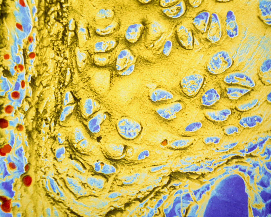
| False-colour scanning electron micrograph (SEM) of human hyaline cartilage from the walls of a bronchus,the main conducting airway of a lung. Hyaline cartilage is found in the walls of conducting airways (the trachea & bronchi) and a specialized type of hyaline cartilage caps the ends of long bones that form joints with other bones. This articular cartilage has a smooth slippery surface which combines with the synovial fluid to ease movement in the joint. This image shows the organization of chondrocytes (cartilage cells) which are embedded in a cartilage matrix that the cells have synthesized. Magnification: x220 at 35mm size | |
| Licence : | Droits gérés |
| Crédit: | Science Photo Library / Photo Insolite Realite |
| Taille de l’image : | 4681 px × 3755 px |
| Model Release : | Non requis |
| Property Release : | Non requis |
| Restrictions : | - |
Prix pour cette image À partir de 45 €
Produit vendu
(Calendrier, Carte postale, Carte de vœux, Impression sur textile, Packaging etc)
À partir de 45 €
Usage commercial
(Affichage, Annonce presse, Annonce TV, Carte, Digital - hors rés. sociaux, Digital - rés. sociaux etc)
À partir de 45 €
Éditorial
(Digital, Journal, Livre, Livre pratique, Magazine, Télévision etc)
À partir de 60 €
Usage non-commercial
(Digital - hors rés. sociaux, Digital - rés. sociaux etc)
À partir de 120 €