Zebrafish muscle
Numéro d’image : 11868447
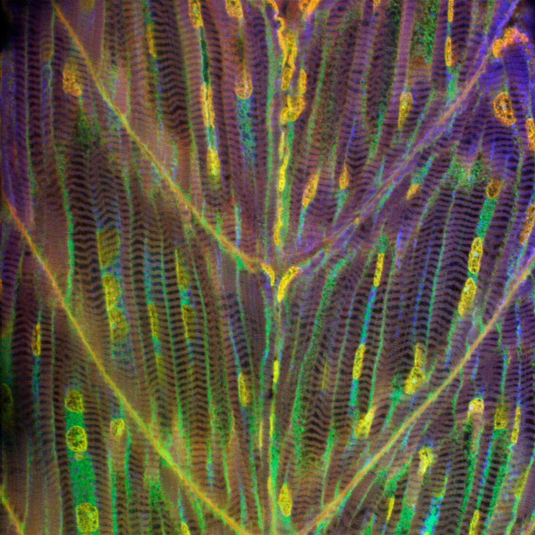
| Zebrafish muscle. Confocal light micrograph of the interior of an intact zebrafish (Danio rerio) larva,showing mytomes (blocks of muscle) in optical section. The tissues have been stained with fluorescent dyes and illuminated with a laser. The long,striated objects are individual muscle cells: cell nuclei are stained yellow,muscle proteins are stained blue/green. A confocal microscope detects light only from the focal point of its objective lens. By moving the focal point,images of thin sections of an intact specimen can be obtained. Magnification: x200 at 6x6cm size | |
| Licence : | Droits gérés |
| Crédit: | Science Photo Library / Reichelt, Stefanie |
| Taille de l’image : | 1024 px × 1024 px |
| Model Release : | Non requis |
| Property Release : | Non requis |
| Restrictions : | - |
Prix pour cette image À partir de 45 €
Produit vendu
(Calendrier, Carte postale, Carte de vœux, Impression sur textile, Packaging etc)
À partir de 45 €
Usage commercial
(Affichage, Annonce presse, Annonce TV, Carte, Digital - hors rés. sociaux, Digital - rés. sociaux etc)
À partir de 45 €
Éditorial
(Digital, Journal, Livre, Livre pratique, Magazine, Télévision etc)
À partir de 60 €
Usage non-commercial
(Digital - hors rés. sociaux, Digital - rés. sociaux etc)
À partir de 120 €
Mots clés
- anatomie,
- animal,
- balayage,
- brachydanio,
- cellules,
- corps,
- DANIO RERIO,
- divisé,
- entiereté,
- fluorescence,
- fluorescent,
- globalité,
- immunofluorescence,
- intact,
- intérieur,
- jeune,
- juvénile,
- larva,
- larvaire,
- larve,
- laser,
- micrographie,
- microscope,
- muscle,
- muscles,
- MYTOME,
- numérisation,
- poisson,
- poisson zèbre,
- poisson-zèbre,
- scanographie,
- tissus,
- totalité,
- transparent