Coloured 3-D CT scan of lower spine
Numéro d’image : 11867720
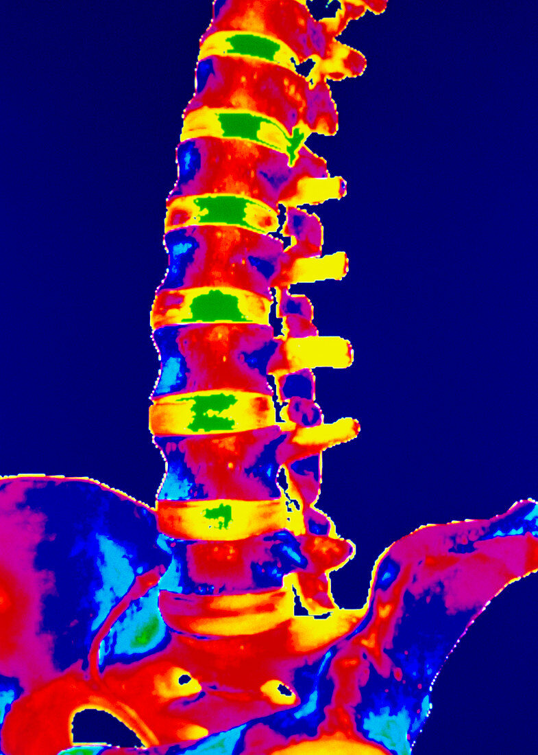
| Lower spine. Coloured three-dimensional computed tomography (CT) scan of the front of the lower spine. The spine is made of a column of 33 cylindrical bones called vertebrae,of which the lower vertebrae (lumbar) are seen here. Between the vertebrae are flexible joints (yellow/green) that both stabilise the spine and allow movement. The projections on the right side of each vertebra provide attachment sites for the ligaments and muscles that hold the spine together. The pelvis is in lower frame. The vertebrae enclose the spinal cord (not seen),a nerve bundle running down from the brain. 3-D CT scans are made by combining multiple X-ray tissue "slices" | |
| Licence : | Droits gérés |
| Crédit: | Science Photo Library / GJLP |
| Taille de l’image : | 3748 px × 5251 px |
| Model Release : | Non requis |
| Property Release : | Non requis |
| Restrictions : | - |
Prix pour cette image À partir de 45 €
Produit vendu
(Calendrier, Carte postale, Carte de vœux, Impression sur textile, Packaging etc)
À partir de 45 €
Usage commercial
(Affichage, Annonce presse, Annonce TV, Carte, Digital - hors rés. sociaux, Digital - rés. sociaux etc)
À partir de 45 €
Éditorial
(Digital, Journal, Livre, Livre pratique, Magazine, Télévision etc)
À partir de 60 €
Usage non-commercial
(Digital - hors rés. sociaux, Digital - rés. sociaux etc)
À partir de 120 €