3-D CT scan of bones of a normal adult pelvis
Numéro d’image : 11867676
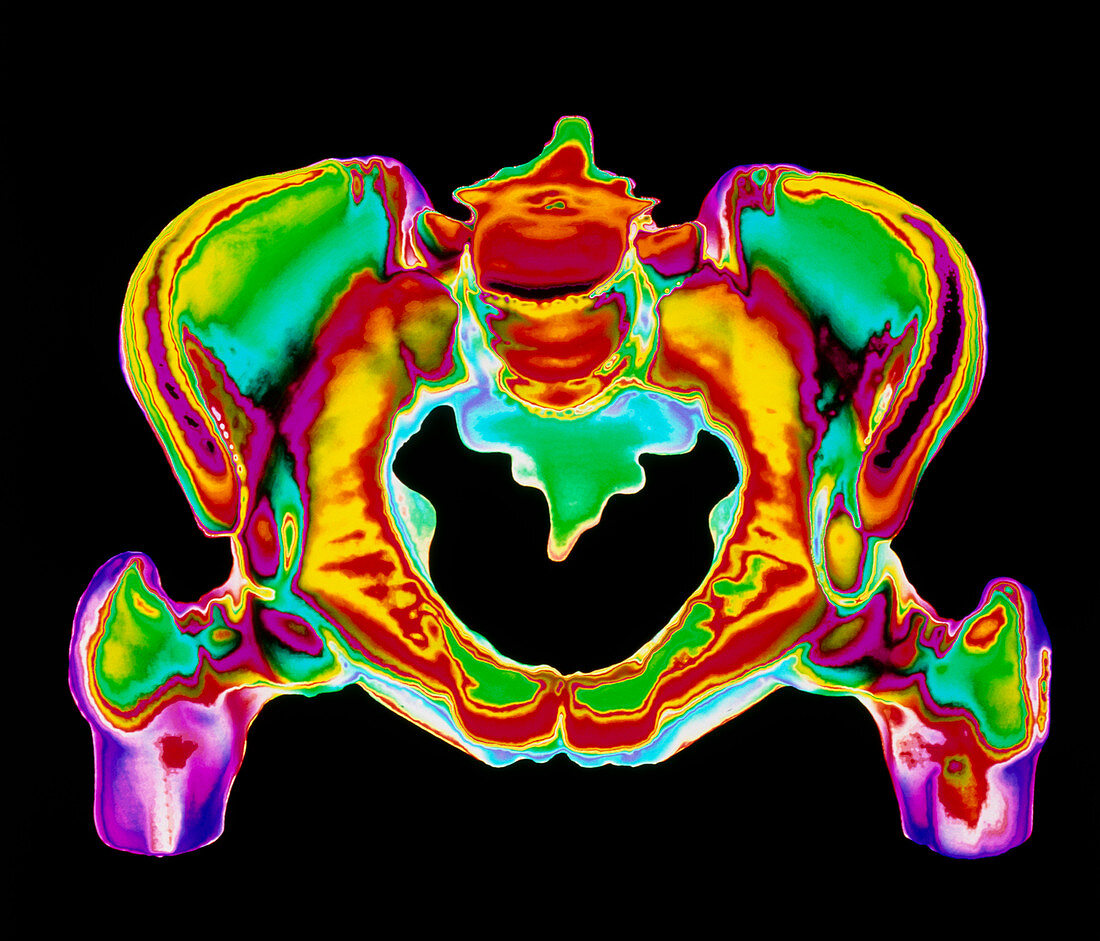
| Human pelvis. Coloured three-dimensional Computed Tomography (CT) scan of bones of a normal adult pelvis. At top centre is the flattened region where lumbar vertebrae articulate with the pelvis; at lower left & right is the rounded head of each femur (thigh bone) which fit into hip sockets of the pelvis. The pelvis itself is formed by bones of the sacrum and coccyx (fused vertebrae,at centre) and two curved innominate or hip bones. The pelvis supports and protects internal organs and tissues of the abdomen,and provides a site for the attachment of muscles of the trunk and lower limbs | |
| Licence : | Droits gérés |
| Crédit: | Science Photo Library |
| Taille de l’image : | 4258 px × 3642 px |
| Model Release : | Non requis |
| Property Release : | Non requis |
| Restrictions : | - |
Prix pour cette image À partir de 45 €
Produit vendu
(Calendrier, Carte postale, Carte de vœux, Impression sur textile, Packaging etc)
À partir de 45 €
Usage commercial
(Affichage, Annonce presse, Annonce TV, Carte, Digital - hors rés. sociaux, Digital - rés. sociaux etc)
À partir de 45 €
Éditorial
(Digital, Journal, Livre, Livre pratique, Magazine, Télévision etc)
À partir de 60 €
Usage non-commercial
(Digital - hors rés. sociaux, Digital - rés. sociaux etc)
À partir de 120 €