Coloured MRI scan of section through a man's hips
Numéro d’image : 11867674
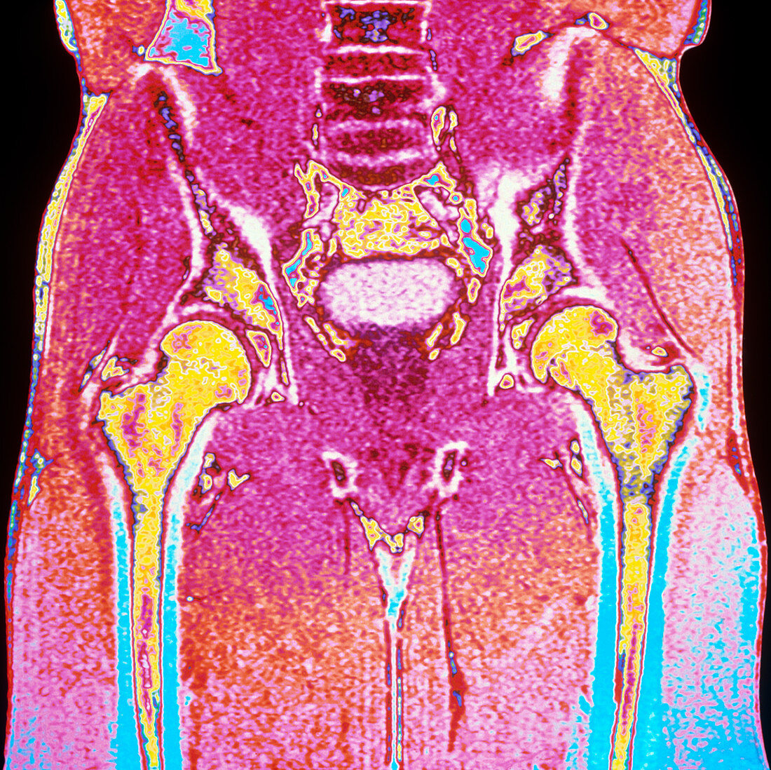
| Man's hips. Coloured Magnetic Resonance Imaging (MRI) scan of a coronal section (front view) through hips of a 29 year old man. At centre top are lumbar vertebrae of the lower spine. The two long femurs (thigh bones,yellow) are clearly seen in each leg; the head of each femur forms a ball joint with partly seen bones of the pelvis. Within the pelvis (at centre) lies the urinary bladder (light pink). Muscle is visible in the hips and upper legs,with pale skin at the surface. The narrow hips of the male contain more muscle than is typical of female hips. MRI scanning uses radio waves to create "slice" images through the body | |
| Licence : | Droits gérés |
| Crédit: | Science Photo Library / Fraser, Simon / Newcastle Upon Tyne / Royal Victoria Infirmary |
| Taille de l’image : | 4252 px × 4246 px |
| Model Release : | Non requis |
| Property Release : | Non requis |
| Restrictions : | - |
Prix pour cette image À partir de 45 €
Produit vendu
(Calendrier, Carte postale, Carte de vœux, Impression sur textile, Packaging etc)
À partir de 45 €
Usage commercial
(Affichage, Annonce presse, Annonce TV, Carte, Digital - hors rés. sociaux, Digital - rés. sociaux etc)
À partir de 45 €
Éditorial
(Digital, Journal, Livre, Livre pratique, Magazine, Télévision etc)
À partir de 60 €
Usage non-commercial
(Digital - hors rés. sociaux, Digital - rés. sociaux etc)
À partir de 120 €