Coloured 3-D CT scan of lower spine
Numéro d’image : 11867653
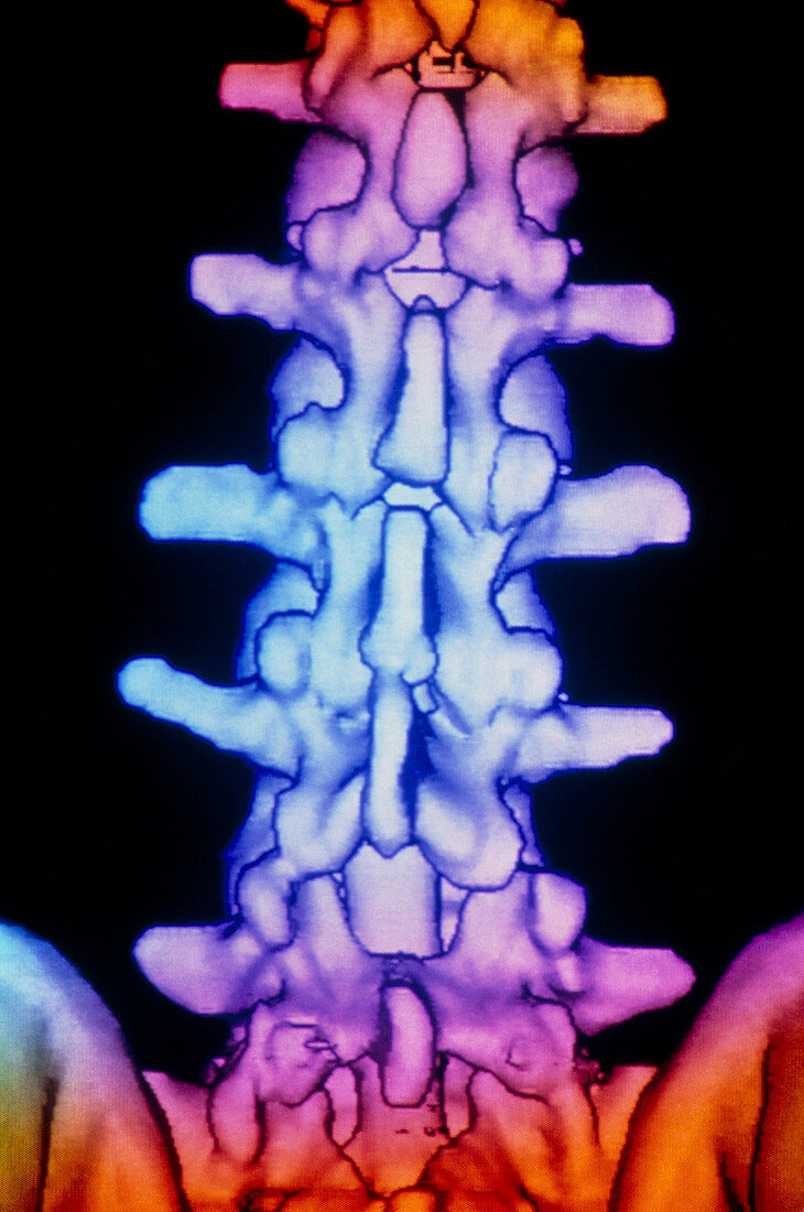
| Lower spine. Coloured 3-D computed tomography (CT) scan of the bones of the lower spine,seen from the rear. The spine is made of a column of 33 cyl- indrical bones called vertebrae,five of which are seen here. Between the vertebrae are flexible joints that both stabilise the spine and allow movement. The bony projections here are behind the cylindrical part of each vertebra,and provide at- tachment sites for the muscles and ligaments that hold the spine together. The pelvis can be seen at lower left and right. Running through the spine is the spinal cord (not seen),a bundle of nerves from the brain. 3-D CT scans are made from multi- ple X-rays combined into one image by a computer | |
| Licence : | Droits gérés |
| Crédit: | Science Photo Library / GCA |
| Taille de l’image : | 2480 px × 3738 px |
| Model Release : | Non requis |
| Property Release : | Non requis |
| Restrictions : | - |
Prix pour cette image À partir de 45 €
Produit vendu
(Calendrier, Carte postale, Carte de vœux, Impression sur textile, Packaging etc)
À partir de 45 €
Usage commercial
(Affichage, Annonce presse, Annonce TV, Carte, Digital - hors rés. sociaux, Digital - rés. sociaux etc)
À partir de 45 €
Éditorial
(Digital, Journal, Livre, Livre pratique, Magazine, Télévision etc)
À partir de 60 €
Usage non-commercial
(Digital - hors rés. sociaux, Digital - rés. sociaux etc)
À partir de 120 €