Coloured X-ray of healthy knee joints,both legs
Numéro d’image : 11867647
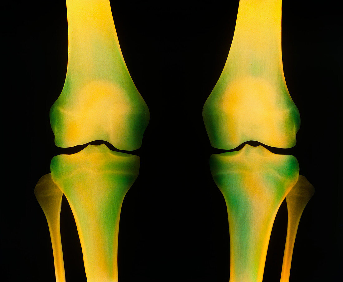
| Normal knees. Coloured X-ray image of healthy knees,showing in each leg the joint between the femur or thigh-bone (top) and tibia (the larger bone at bottom). This is a frontal view of both legs of a fifteen-year-old girl. The patella,also known as the kneecap,can be seen as an oval yellow area in the region of the head of each femur. Two discs (unseen) of cartilage,the menisci,cover the end of each femur and tibia. These discs reduce the friction between the bones during movement and increase the stability of the knee. The fibula is the thin long bone of the lower leg (bottom,far left and far right) and is not part of the knee joint articulation | |
| Licence : | Droits gérés |
| Crédit: | Science Photo Library |
| Taille de l’image : | 5023 px × 4137 px |
| Model Release : | Non requis |
| Property Release : | Non requis |
| Restrictions : | - |
Prix pour cette image À partir de 45 €
Produit vendu
(Calendrier, Carte postale, Carte de vœux, Impression sur textile, Packaging etc)
À partir de 45 €
Usage commercial
(Affichage, Annonce presse, Annonce TV, Carte, Digital - hors rés. sociaux, Digital - rés. sociaux etc)
À partir de 45 €
Éditorial
(Digital, Journal, Livre, Livre pratique, Magazine, Télévision etc)
À partir de 60 €
Usage non-commercial
(Digital - hors rés. sociaux, Digital - rés. sociaux etc)
À partir de 120 €