Fossilised compact bone,SEM
Numéro d’image : 11867531
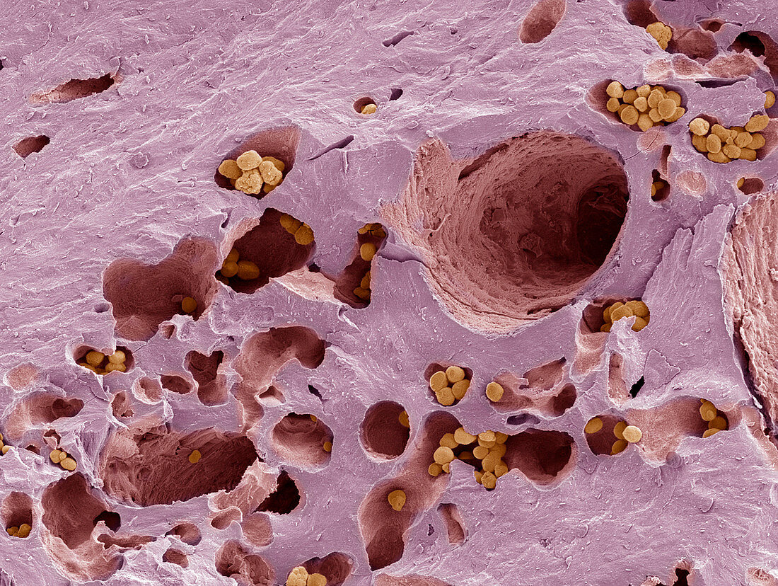
| Fossilised compact bone. Coloured scanning electron micrograph (SEM) of a section through fossilised compact bone. This tissue is found in the dense walls of the shafts of bones. It consists of concentric layers of collagen- containing matrix (lamellae,pink),around Haversian canals (large holes),which contain blood and lymph vessels,and nerves. These canals run the length of the bone. The lamellae and canals together form a Haversian system,or osteon. Many systems are arranged in columns running parallel to the long axis of the bone. Mineral deposits (yellow) have collected in the lacunae (smaller holes) of the bone during fossilisation | |
| Licence : | Droits gérés |
| Crédit: | Science Photo Library / Gschmeissner, Steve |
| Taille de l’image : | 3405 px × 2564 px |
| Model Release : | Non requis |
| Property Release : | Non requis |
| Restrictions : | - |
Prix pour cette image À partir de 45 €
Produit vendu
(Calendrier, Carte postale, Carte de vœux, Impression sur textile, Packaging etc)
À partir de 45 €
Usage commercial
(Affichage, Annonce presse, Annonce TV, Carte, Digital - hors rés. sociaux, Digital - rés. sociaux etc)
À partir de 45 €
Éditorial
(Digital, Journal, Livre, Livre pratique, Magazine, Télévision etc)
À partir de 60 €
Usage non-commercial
(Digital - hors rés. sociaux, Digital - rés. sociaux etc)
À partir de 120 €
Mots clés
- agrandissement,
- anatomie,
- anatomique,
- biologie,
- biologique,
- canal,
- canaux,
- catégorie,
- cellule,
- collagène,
- coloré,
- colorié,
- colorisé,
- couche concentrique,
- coupe,
- divisé,
- en bonne santé,
- fossile,
- images,
- Lacunae,
- Lacune,
- lamella,
- lamelle,
- M.E.B.,
- matrice,
- MEB,
- microscope électronique à balayage,
- normal,
- os compact,
- ostéocyte,
- ostéon,
- ostéons,
- partie,
- rose,
- sain,
- section,
- sujets microscopiques,
- système de Havers,
- tissus