Compact bone
Numéro d’image : 11867471
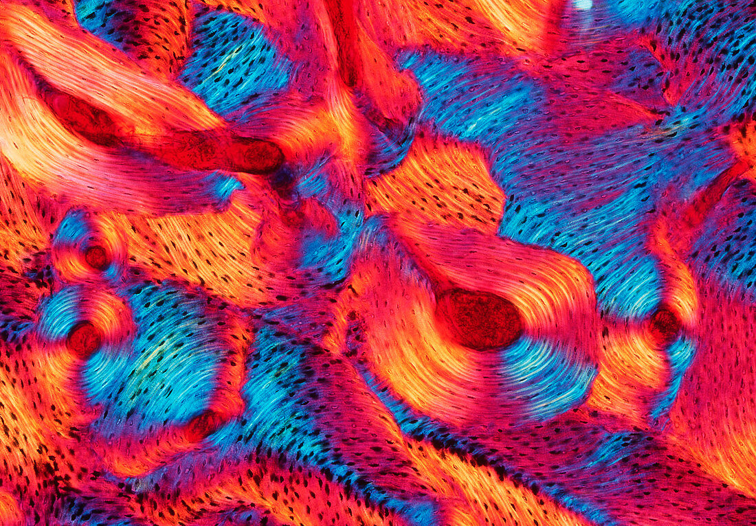
| Compact bone. Polarized light micrograph (PLM) of a section through human compact bone. This tissue is found in the dense walls of the shafts of bones. It forms concentric layers (lamellae,blue,purple and orange) around Haversian canals (red circles),which contain blood,lymph vessels and nerves. The lamellae and canals together form a Haversian system. Many Haversian systems are arranged in columns running parallel to the long axis of the bone. Osteocytes,the cells responsible for the maintenance of the bony matrix,are housed in spaces known as lacunae (small black dots). Magnification: x50 at 6x7cm size x25 at 35mm size | |
| Licence : | Droits gérés |
| Crédit: | Science Photo Library / Innerspace Imaging |
| Taille de l’image : | 4500 px × 3134 px |
| Model Release : | Non requis |
| Property Release : | Non requis |
| Restrictions : | - |
Prix pour cette image À partir de 45 €
Produit vendu
(Calendrier, Carte postale, Carte de vœux, Impression sur textile, Packaging etc)
À partir de 45 €
Usage commercial
(Affichage, Annonce presse, Annonce TV, Carte, Digital - hors rés. sociaux, Digital - rés. sociaux etc)
À partir de 45 €
Éditorial
(Digital, Journal, Livre, Livre pratique, Magazine, Télévision etc)
À partir de 60 €
Usage non-commercial
(Digital - hors rés. sociaux, Digital - rés. sociaux etc)
À partir de 120 €