Foetal bone
Numéro d’image : 11867467
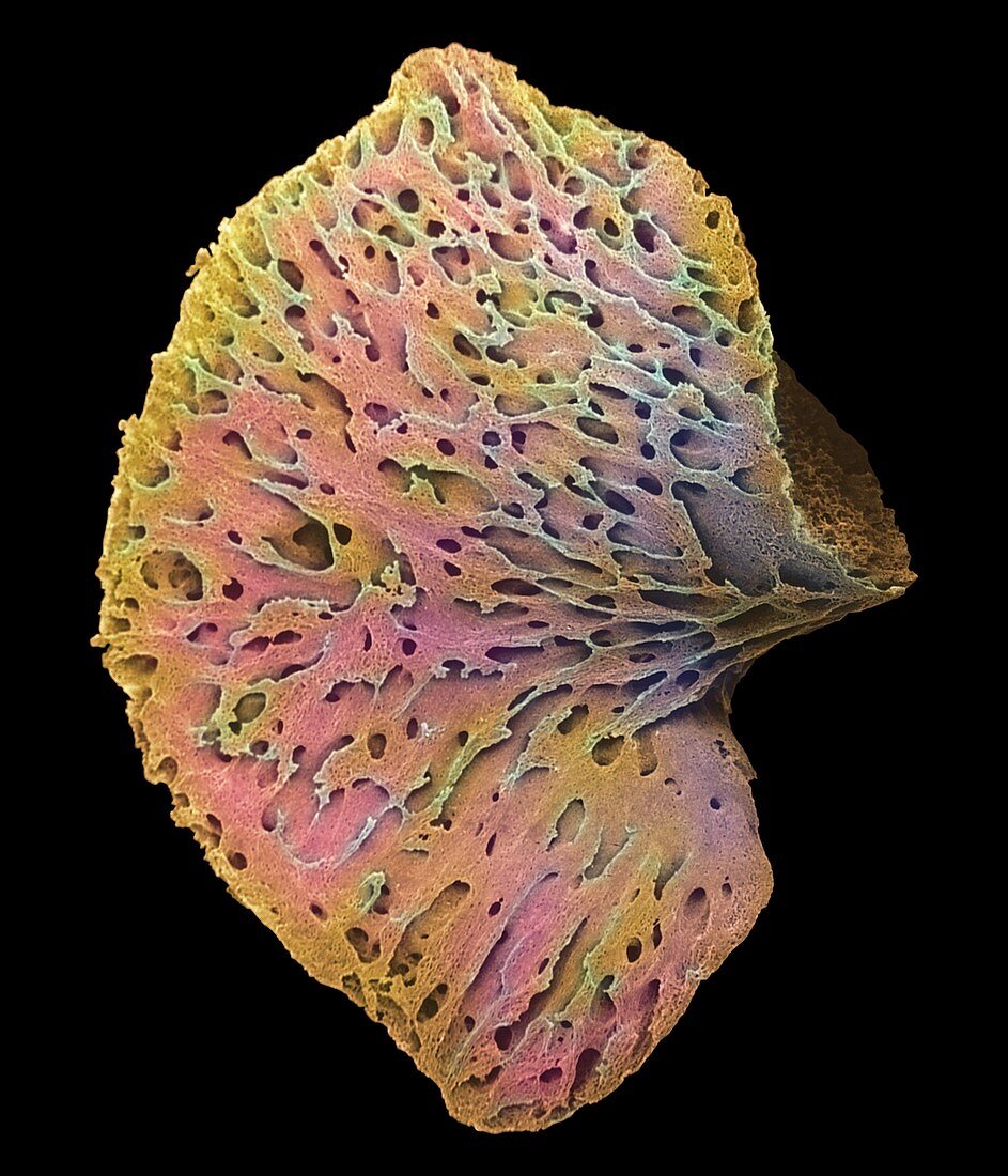
| Foetal bone. Coloured scanning electron micrograph (SEM) of a pelvic bone of an 11 week old foetus. Only the calcified matrix of the bone remains as the cartilage has been lost during the processing of the sample. The pelvis is initially formed of cartilage in the foetus,but it is steadily replaced by bone as the foetus grows. Some cartilage is still present in childrens' bones. Magnification: x12.5 at 6x7cm size | |
| Licence : | Droits gérés |
| Crédit: | Science Photo Library / Moscoso, Dr. G. |
| Taille de l’image : | 2400 px × 2800 px |
| Model Release : | Non requis |
| Property Release : | Non requis |
| Restrictions : | - |
Prix pour cette image À partir de 45 €
Produit vendu
(Calendrier, Carte postale, Carte de vœux, Impression sur textile, Packaging etc)
À partir de 45 €
Usage commercial
(Affichage, Annonce presse, Annonce TV, Carte, Digital - hors rés. sociaux, Digital - rés. sociaux etc)
À partir de 45 €
Éditorial
(Digital, Journal, Livre, Livre pratique, Magazine, Télévision etc)
À partir de 60 €
Usage non-commercial
(Digital - hors rés. sociaux, Digital - rés. sociaux etc)
À partir de 120 €