Foetus compact bone
Numéro d’image : 11867465
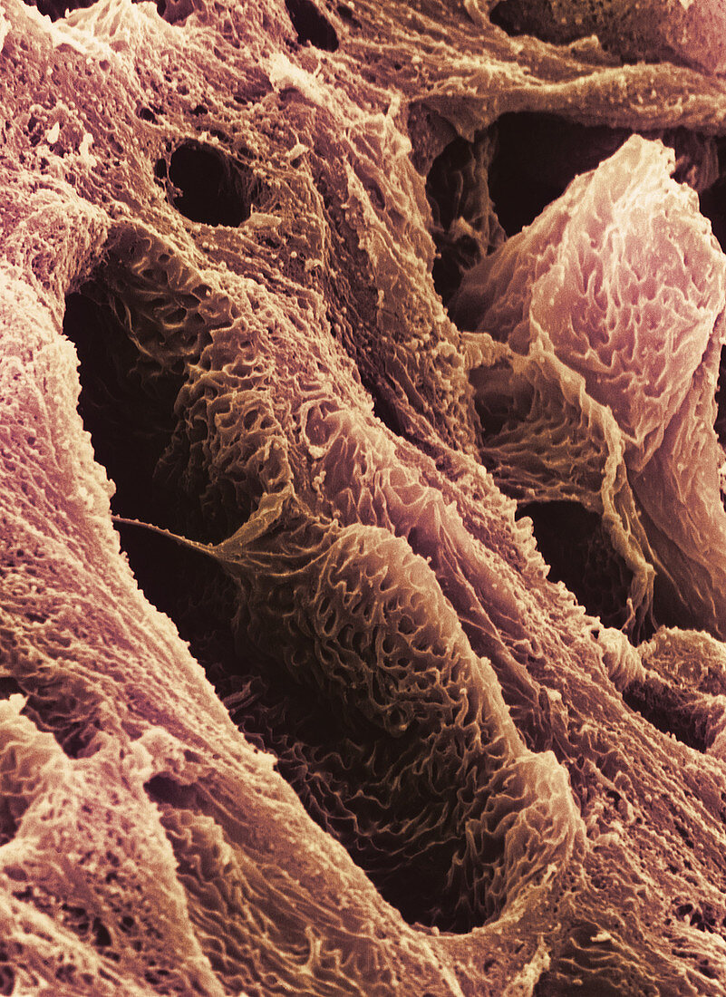
| Foetus compact bone. Coloured scanning electron micrograph (SEM) of a section through the develop- ing bone in the foot of a human foetus. This region,known as the epiphysis,is at the end of the long bone within a foetal foot. It will develop separately from the main shaft of the bone,but will eventually fuse to form a complete bone. The dark areas at lower left & upper right are Haversian canals,an interconnecting neurova- scular network containing blood vessels and nerves. The small cavities on the bone's surface are canaliculi. These are interconnecting canals which allow circulation of tissue fluid and the circulation of metabolites. Magnification unknown | |
| Licence : | Droits gérés |
| Crédit: | Science Photo Library / Gschmeissner, Steve |
| Taille de l’image : | 1922 px × 2640 px |
| Model Release : | Non requis |
| Property Release : | Non requis |
| Restrictions : | - |
Prix pour cette image À partir de 45 €
Produit vendu
(Calendrier, Carte postale, Carte de vœux, Impression sur textile, Packaging etc)
À partir de 45 €
Usage commercial
(Affichage, Annonce presse, Annonce TV, Carte, Digital - hors rés. sociaux, Digital - rés. sociaux etc)
À partir de 45 €
Éditorial
(Digital, Journal, Livre, Livre pratique, Magazine, Télévision etc)
À partir de 60 €
Usage non-commercial
(Digital - hors rés. sociaux, Digital - rés. sociaux etc)
À partir de 120 €