LM of compact bone,showing a Haversian system
Numéro d’image : 11867418
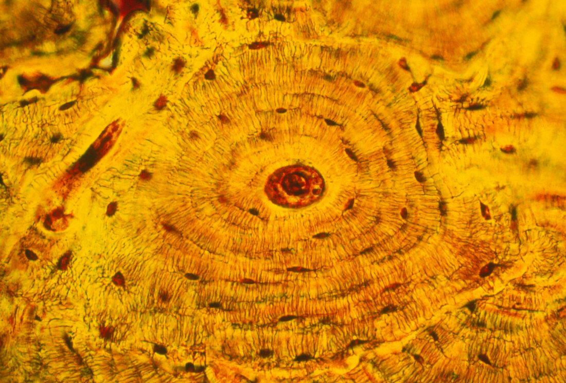
| Light micrograph of human compact bone,which comprises the dense walls of the shafts of long bones. The image shows the organization of compact bone into concentric bony layers (lamellae) arranged around channels containing blood,lymph vessels and nerves (centre). The lamellae and channel form an Haversian system,a number of which appear here in various sections. The Haversian systems are arranged in columns which run parallel to the long axes of the bone. The small dark spots are spaces (lacunae) which house osteocytes,the cells responsible for the maintenance of the bony matrix. Magnification: x320 at 6x4.5cm size,x203 at 35mm size | |
| Licence : | Droits gérés |
| Crédit: | Science Photo Library / Burbidge, John |
| Taille de l’image : | 3667 px × 2480 px |
| Model Release : | Non requis |
| Property Release : | Non requis |
| Restrictions : | - |
Prix pour cette image À partir de 45 €
Produit vendu
(Calendrier, Carte postale, Carte de vœux, Impression sur textile, Packaging etc)
À partir de 45 €
Usage commercial
(Affichage, Annonce presse, Annonce TV, Carte, Digital - hors rés. sociaux, Digital - rés. sociaux etc)
À partir de 45 €
Éditorial
(Digital, Journal, Livre, Livre pratique, Magazine, Télévision etc)
À partir de 60 €
Usage non-commercial
(Digital - hors rés. sociaux, Digital - rés. sociaux etc)
À partir de 120 €