Pregnancy,16th century artwork
Numéro d’image : 11866582
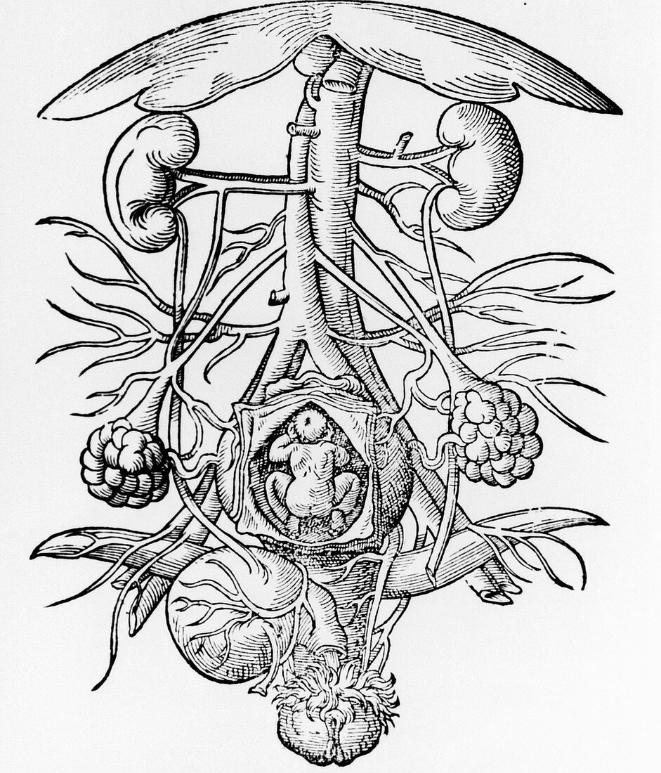
| Anatomy of pregnancy. Engraving from the 16th cen- tury of the anatomy of a pregnant woman's urogeni- tal system. Within the opened uterus (lower cent- re) is a foetus. The ovaries are at lower left & lower right,with the vagina at bottom centre. At bottom left is the bladder,whilst the kidneys are at upper left and upper right. The vertical blood vessels between the kidneys are the aorta artery (narrower of the 2) and the inferior vena cava vein. These split (at centre) to form iliac veins & arteries. The umbrella-shaped object (at top) is the liver. Image taken from De conceptu et generatione hominis (1580) by Jakob Rueff | |
| Licence : | Droits gérés |
| Crédit: | Science Photo Library / National Library of Medicine |
| Taille de l’image : | 3898 px × 4556 px |
| Model Release : | Non requis |
| Property Release : | Non requis |
| Restrictions : | - |
Prix pour cette image À partir de 45 €
Produit vendu
(Calendrier, Carte postale, Carte de vœux, Impression sur textile, Packaging etc)
À partir de 45 €
Usage commercial
(Affichage, Annonce presse, Annonce TV, Carte, Digital - hors rés. sociaux, Digital - rés. sociaux etc)
À partir de 45 €
Éditorial
(Digital, Journal, Livre, Livre pratique, Magazine, Télévision etc)
À partir de 60 €
Usage non-commercial
(Digital - hors rés. sociaux, Digital - rés. sociaux etc)
À partir de 120 €