Artwork of a developing human embryo
Numéro d’image : 11866581
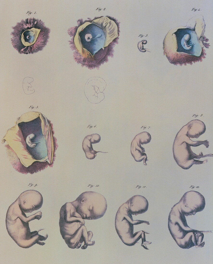
| Human embryo. Composite artwork showing 12 images of a developing human embryo. Four images (1,2,4 & 5) show the embryo inside the womb. The embryos in Figures 1 and 2 have a larger line drawing beneath them to show more detail. The long struct- ure attached to the belly of the embryos is the umbilical cord which delivers food and oxygen to the embryo from the mother. In the early stages of development,different areas of the embryo become differentiated to form the various struct- ures and organs. In later stages these structures develop,making the embryo more recognisable as a human. Image taken from Manuel D'Anatomie de Corps Humain (1825) by Jules Cloquet. Age of different stages unknown | |
| Licence : | Droits gérés |
| Crédit: | Science Photo Library / Charmet, Jean-Loup |
| Taille de l’image : | 2854 px × 3543 px |
| Model Release : | Non requis |
| Property Release : | Non requis |
| Restrictions : | - |
Prix pour cette image À partir de 45 €
Produit vendu
(Calendrier, Carte postale, Carte de vœux, Impression sur textile, Packaging etc)
À partir de 45 €
Usage commercial
(Affichage, Annonce presse, Annonce TV, Carte, Digital - hors rés. sociaux, Digital - rés. sociaux etc)
À partir de 45 €
Éditorial
(Digital, Journal, Livre, Livre pratique, Magazine, Télévision etc)
À partir de 60 €
Usage non-commercial
(Digital - hors rés. sociaux, Digital - rés. sociaux etc)
À partir de 120 €