Pathologist using LM to examine bone biopsy slide
Numéro d’image : 11864568
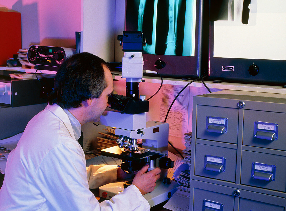
| MODEL RELEASED. Hospital pathologist using a light microscope to examine a bone biopsy section from the tibia (shin bone) of a man aged 19 years. The left pair of X-ray films (blue) reveal a tumour,possibly cancerous,of the tibia (dark areas towards bottom). A small section of bone,removed during exploratory surgery,is embedded in resin and cut into thin slices. After staining and mounting on a slide,the sample is carefully examined to establish an accurate pathological diagnosis. The X-ray film on the right shows the knee of another patient. Model: Archie Malcolm | |
| Licence : | Droits gérés |
| Crédit: | Science Photo Library / RVI, NEWCASTLE / UNIVERSITY PATHOLOGY DEPT. / SIMON FRASER |
| Taille de l’image : | 4795 px × 3550 px |
| Model Release : | Disponible |
| Property Release : | Non requis |
| Restrictions : | - |
Prix pour cette image À partir de 45 €
Produit vendu
(Calendrier, Carte postale, Carte de vœux, Impression sur textile, Packaging etc)
À partir de 45 €
Usage commercial
(Affichage, Annonce presse, Annonce TV, Carte, Digital - hors rés. sociaux, Digital - rés. sociaux etc)
À partir de 45 €
Éditorial
(Digital, Journal, Livre, Livre pratique, Magazine, Télévision etc)
À partir de 60 €
Usage non-commercial
(Digital - hors rés. sociaux, Digital - rés. sociaux etc)
À partir de 120 €