Coloured PET scan of brain during drugged sleep
Numéro d’image : 11863978
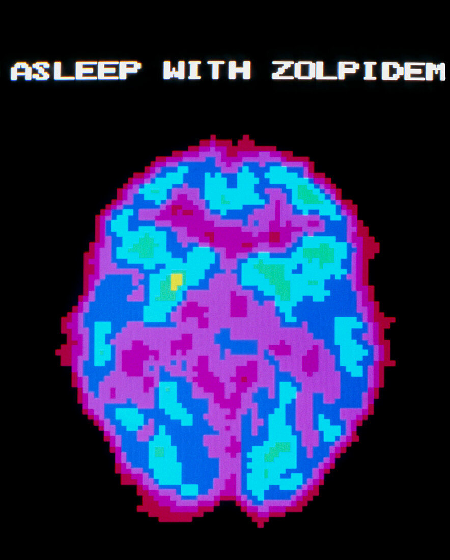
| Brain during drugged sleep. Coloured Positron Emission Tomography (PET) scan of the human brain during drugged sleep. The drug was zolpidem. The colour-coding depicts active cerebral brain areas (yellow) through to inactive areas (purple). The brain is inactive,undergoing a deeper sleep phase than normal NREM sleep. Sleeping drugs promote sleep by reducing nerve cell activity in the brain. PET scanning shows the metabolic activity of the brain. A radioactive tracer (here,radio- labelled glucose) is injected into the patient's blood and absorbed by active tissues of the brain. The PET scanner detects photons emitted by the tracer,to produce a "slice" image of the brain | |
| Licence : | Droits gérés |
| Crédit: | Science Photo Library / Morgan, Hank |
| Taille de l’image : | 3579 px × 4455 px |
| Model Release : | Non requis |
| Property Release : | Non requis |
| Restrictions : |
|
Prix pour cette image À partir de 45 €
Produit vendu
(Calendrier, Carte postale, Carte de vœux, Impression sur textile, Packaging etc)
À partir de 45 €
Usage commercial
(Affichage, Annonce presse, Annonce TV, Carte, Digital - hors rés. sociaux, Digital - rés. sociaux etc)
À partir de 45 €
Éditorial
(Digital, Journal, Livre, Livre pratique, Magazine, Télévision etc)
À partir de 60 €
Usage non-commercial
(Digital - hors rés. sociaux, Digital - rés. sociaux etc)
À partir de 120 €