Brain in REM sleep
Numéro d’image : 11863974
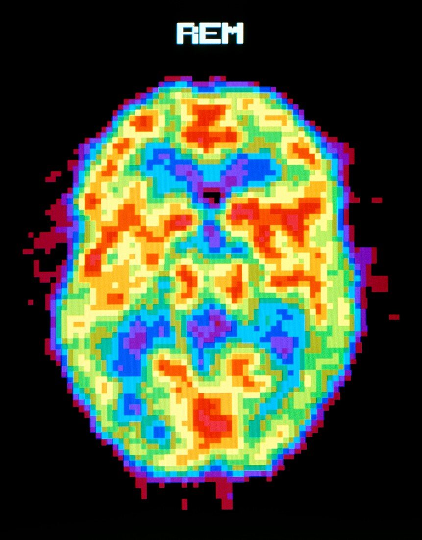
| Brain during REM sleep. Coloured Positron Emission Tomography (PET) scan of the human brain during REM (rapid eye movement) sleep. Colour-coding depicts active cerebral brain areas (red) through to inactive areas (blue). During the REM sleep phase,the brain is active and dreaming,showing similar activity when awake. In the non-REM phase of sleep the brain is in a deeper,less active sleep. PET scanning shows metabolic activity of the brain. A radioactive tracer (here,radio- labelled glucose) is injected into the bloodstream and absorbed by active tissues of the brain. The PET scanner detects photons emitted by the tracer,to produce a "slice" image of the brain | |
| Licence : | Droits gérés |
| Crédit: | Science Photo Library / Morgan, Hank |
| Taille de l’image : | 3736 px × 4772 px |
| Model Release : | Non requis |
| Property Release : | Non requis |
| Restrictions : |
|
Prix pour cette image À partir de 45 €
Produit vendu
(Calendrier, Carte postale, Carte de vœux, Impression sur textile, Packaging etc)
À partir de 45 €
Usage commercial
(Affichage, Annonce presse, Annonce TV, Carte, Digital - hors rés. sociaux, Digital - rés. sociaux etc)
À partir de 45 €
Éditorial
(Digital, Journal, Livre, Livre pratique, Magazine, Télévision etc)
À partir de 60 €
Usage non-commercial
(Digital - hors rés. sociaux, Digital - rés. sociaux etc)
À partir de 120 €
Mots clés
- activité,
- cerveau,
- corps,
- coupe longitudinale,
- médecine,
- médical,
- médicale,
- mesure physiologique,
- mouvement oculaire rapide,
- neuro-imagerie,
- neuroimagerie,
- pet scan,
- physiologie,
- psychologie,
- recherche sur le sommeil,
- scanner du cerveau,
- soins de santé,
- sommeil,
- T.E.P.,
- TEP,
- test,
- tester,
- tomographie à émission de positrons,
- tomographie par émission de positrons