PET scan of a brain during normal sleep
Numéro d’image : 11863973
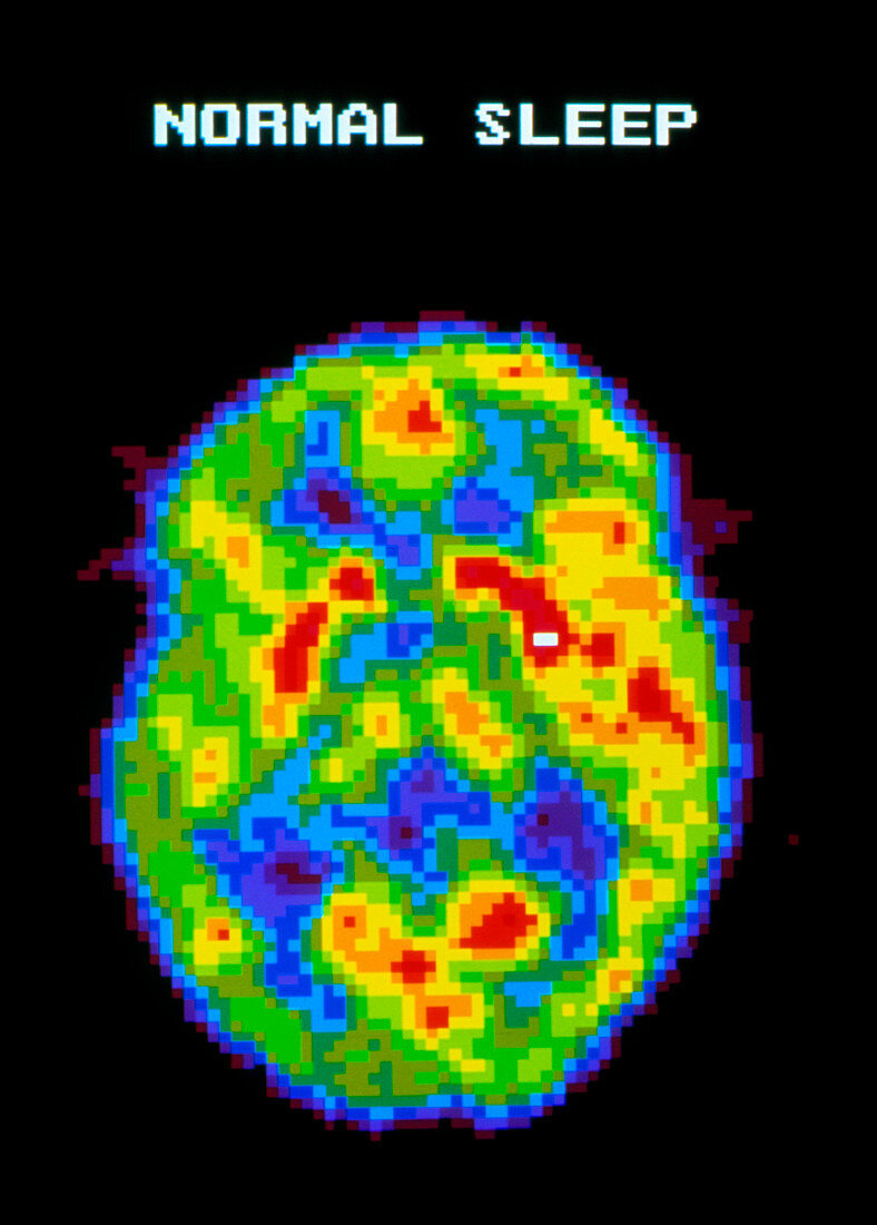
| Brain during normal sleep. Coloured Positron Emission Tomography (PET) scan of the human brain during normal sleep. Colour-coding depicts active cerebral brain areas (red) through to inactive areas (blue). During normal sleep the brain is slightly active,less active than when awake,but more active than in deep sleep. Brain activity increases further during REM (rapid eye movement) "dreaming" sleep. PET scanning shows metabolic activity of the brain. A radioactive tracer (here,radio-labelled glucose) is injected into the blood and absorbed by active tissues of the brain. The PET scanner detects photons emitted by the tracer,to produce a "slice" image of the brain | |
| Licence : | Droits gérés |
| Crédit: | Science Photo Library / Morgan, Hank |
| Taille de l’image : | 2538 px × 3543 px |
| Model Release : | Non requis |
| Property Release : | Non requis |
| Restrictions : |
|
Prix pour cette image À partir de 45 €
Produit vendu
(Calendrier, Carte postale, Carte de vœux, Impression sur textile, Packaging etc)
À partir de 45 €
Usage commercial
(Affichage, Annonce presse, Annonce TV, Carte, Digital - hors rés. sociaux, Digital - rés. sociaux etc)
À partir de 45 €
Éditorial
(Digital, Journal, Livre, Livre pratique, Magazine, Télévision etc)
À partir de 60 €
Usage non-commercial
(Digital - hors rés. sociaux, Digital - rés. sociaux etc)
À partir de 120 €