Testicular cancer
Numéro d’image : 11863647
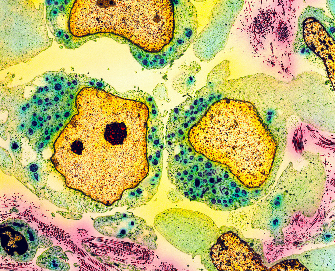
| Testicular cancer. Coloured transmission electron micrograph (TEM) of a section through teratoma cancer cells in a testis. Three rapidly-dividing cancer cells are seen at centre left,centre right and lower right. They have large,irregular nuclei (pale brown) and green cytoplasm. Malignant tera- toma of the testis mostly affects young men. They are thought to develop from cells misplaced during embryonic development. Diagnosis is confirmed by removal of the testis and microscopic examination. This may effect a cure,but radiotherapy and anticancer drugs often follow to stop the cancer spreading through the body. 95-100% of cases detected early are cured. Magnification unknown | |
| Licence : | Droits gérés |
| Crédit: | Science Photo Library / Gschmeissner, Steve |
| Taille de l’image : | 3543 px × 2869 px |
| Model Release : | Non requis |
| Property Release : | Non requis |
| Restrictions : | - |
Prix pour cette image À partir de 45 €
Produit vendu
(Calendrier, Carte postale, Carte de vœux, Impression sur textile, Packaging etc)
À partir de 45 €
Usage commercial
(Affichage, Annonce presse, Annonce TV, Carte, Digital - hors rés. sociaux, Digital - rés. sociaux etc)
À partir de 45 €
Éditorial
(Digital, Journal, Livre, Livre pratique, Magazine, Télévision etc)
À partir de 60 €
Usage non-commercial
(Digital - hors rés. sociaux, Digital - rés. sociaux etc)
À partir de 120 €