LM showing benign prostatic hyperplasia
Numéro d’image : 11863592
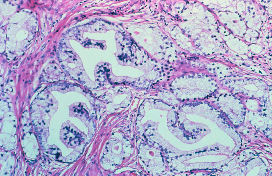
| Light micrograph showing benign prostatic hyperplasia. Hyperplasia describes an increase in cell numbers due to cell division. In the prostate gland,the inner portion & its surrounding fibromuscular stroma may become hyperplastic. Here,the acini (white spaces) are lined with tall,purple-stained cells with small basal nuclei; the glandular lining appears buckled into papillary folds. Adjacent acini are separated by thickened fibromuscular connective tissue (red stain). Since the inner (paraurethral) part of the gland is involved,compression of the urethra is common. This accounts for the symptoms of urinary retention that are common in elderly men | |
| Licence : | Droits gérés |
| Crédit: | Science Photo Library / Biophoto Associates |
| Taille de l’image : | 3643 px × 2362 px |
| Model Release : | Non requis |
| Property Release : | Non requis |
| Restrictions : | - |
Prix pour cette image À partir de 45 €
Produit vendu
(Calendrier, Carte postale, Carte de vœux, Impression sur textile, Packaging etc)
À partir de 45 €
Usage commercial
(Affichage, Annonce presse, Annonce TV, Carte, Digital - hors rés. sociaux, Digital - rés. sociaux etc)
À partir de 45 €
Éditorial
(Digital, Journal, Livre, Livre pratique, Magazine, Télévision etc)
À partir de 60 €
Usage non-commercial
(Digital - hors rés. sociaux, Digital - rés. sociaux etc)
À partir de 120 €