Light micrograph of normal cells in cervical smear
Numéro d’image : 11862734
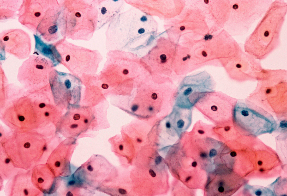
| Cervical cells. Light micrograph of normal squamous epithelial cells in a cervical smear. These cells make up the outer wall of the cervix,the passage linking the uterus to the vagina. The cells have large amounts of cytoplasm (stained pink or blue) and small nuclei (black dots). Cervical smears are examined to alert a doctor to the cellular changes associated with the development of cervical infection or cancer. Papanicolaou stained. Magnification: x400 at 35mm x670 at 5x7cm size | |
| Licence : | Droits gérés |
| Crédit: | Science Photo Library / Walker, Dr. E. |
| Taille de l’image : | 4425 px × 3025 px |
| Model Release : | Non requis |
| Property Release : | Non requis |
| Restrictions : | - |
Prix pour cette image À partir de 45 €
Produit vendu
(Calendrier, Carte postale, Carte de vœux, Impression sur textile, Packaging etc)
À partir de 45 €
Usage commercial
(Affichage, Annonce presse, Annonce TV, Carte, Digital - hors rés. sociaux, Digital - rés. sociaux etc)
À partir de 45 €
Éditorial
(Digital, Journal, Livre, Livre pratique, Magazine, Télévision etc)
À partir de 60 €
Usage non-commercial
(Digital - hors rés. sociaux, Digital - rés. sociaux etc)
À partir de 120 €
Mots clés
- col de l'utérus,
- dénigrement,
- désordre,
- diffamation,
- epithelium,
- épithélium,
- épithélium malphigien,
- état,
- frottis,
- frottis cervical,
- gynécologie,
- gynécologique,
- maladie,
- médecine,
- médical,
- médicale,
- normal,
- papanicolaou,
- pathologie de la reproduction,
- soins de santé,
- squameux,
- système reproducteur féminin,
- tache,
- trouble