Coloured CT scan of large fibroid tumour in uterus
Numéro d’image : 11862678
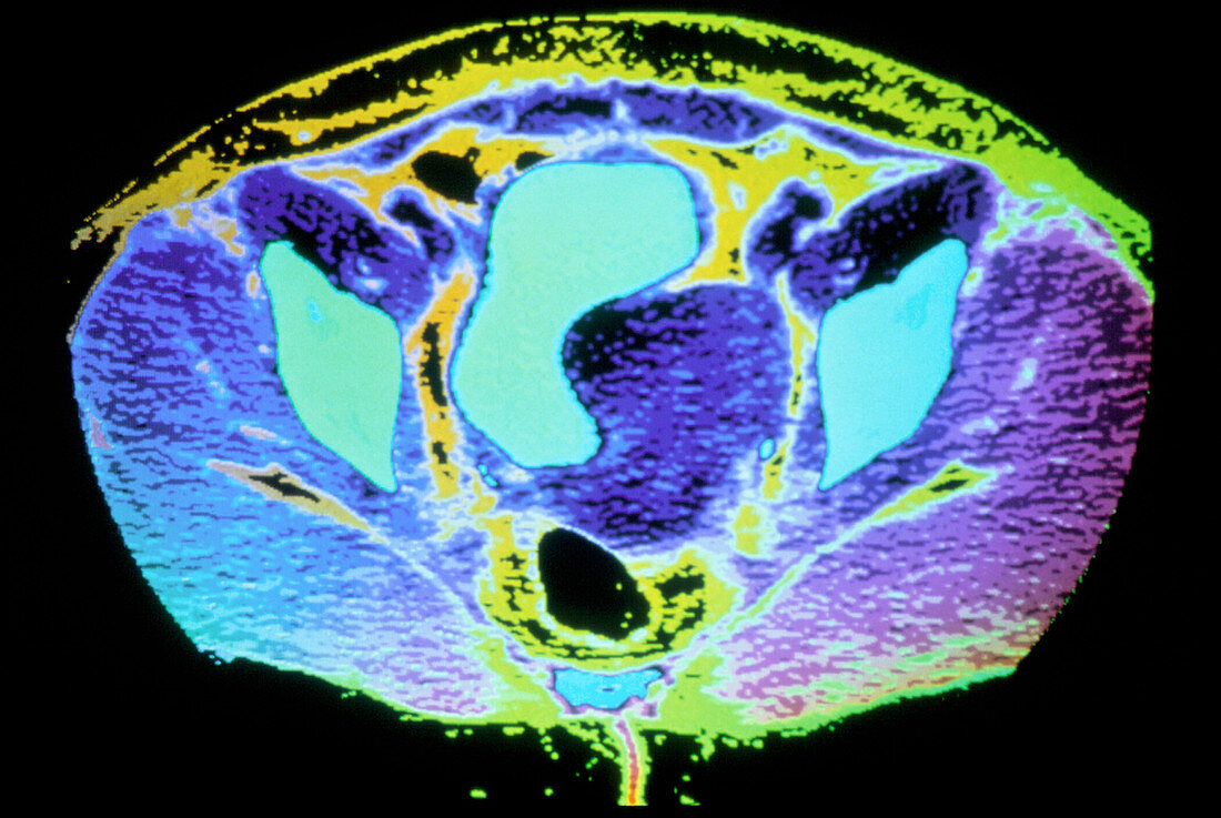
| Fibroid in uterus. Coloured computed tomography scan of a section through a woman's lower abdomen showing a large fibroid tumour pressing against the bladder. The fibroid is the dark blue body in the centre. Above it is the bladder (light blue). To its left and right are bones of the pelvis (also light blue). The black circle at the bottom is the lumen of the uterus,with the rest of the uterine wall around it yellow. Fibroids are benign connective tissue tumours of the uterus. They occur in about 20% of women over age 30,and vary from pea-size to as big as a cauliflower. Small fibroids are harmless,but very large ones may in- terfere with nearby organs,as has happened here | |
| Licence : | Droits gérés |
| Crédit: | Science Photo Library / GJLP |
| Taille de l’image : | 2300 px × 1540 px |
| Model Release : | Non requis |
| Property Release : | Non requis |
| Restrictions : | - |
Prix pour cette image À partir de 45 €
Produit vendu
(Calendrier, Carte postale, Carte de vœux, Impression sur textile, Packaging etc)
À partir de 45 €
Usage commercial
(Affichage, Annonce presse, Annonce TV, Carte, Digital - hors rés. sociaux, Digital - rés. sociaux etc)
À partir de 45 €
Éditorial
(Digital, Journal, Livre, Livre pratique, Magazine, Télévision etc)
À partir de 60 €
Usage non-commercial
(Digital - hors rés. sociaux, Digital - rés. sociaux etc)
À partir de 120 €