Coloured SEM of Sertoli-Leydig tumour in ovary
Numéro d’image : 11862670
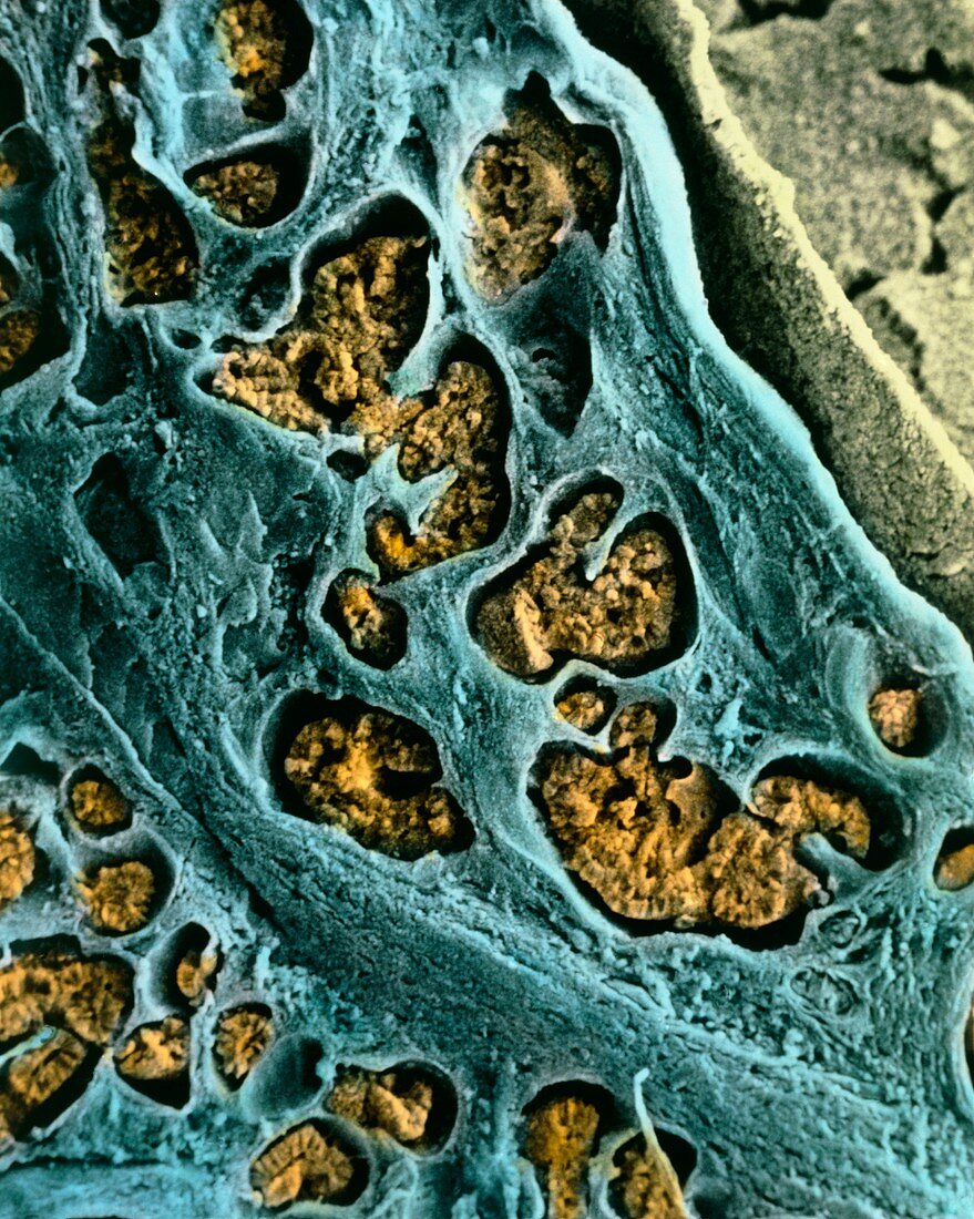
| Sertoli-Leydig tumour in ovary. Coloured Scanning Electron Micrograph (SEM) of a section through a Sertoli-Leydig tumour in the human ovary. Known also as an "arrhenoblastoma" or "androblastoma",up to 30% of these tumours are malignant (cancer- ous). Here,epithelial cells (orange) form branching cords. These epithelial components are contained in tubules (blue). Sertoli cells are the epithelial component,and Leydig cells are the interstitial component between tubules (as at lower right). A Sertoli-Leydig tumour can produce hormones,and may cause masculinization in women. Magnification: x40 at 6x7cm size. x50 at 4x5 | |
| Licence : | Droits gérés |
| Crédit: | Science Photo Library / PROFESSORS P.M. MOTTA & S. MAKABE |
| Taille de l’image : | 3760 px × 4705 px |
| Model Release : | Non requis |
| Property Release : | Non requis |
| Restrictions : | - |
Prix pour cette image À partir de 45 €
Produit vendu
(Calendrier, Carte postale, Carte de vœux, Impression sur textile, Packaging etc)
À partir de 45 €
Usage commercial
(Affichage, Annonce presse, Annonce TV, Carte, Digital - hors rés. sociaux, Digital - rés. sociaux etc)
À partir de 45 €
Éditorial
(Digital, Journal, Livre, Livre pratique, Magazine, Télévision etc)
À partir de 60 €
Usage non-commercial
(Digital - hors rés. sociaux, Digital - rés. sociaux etc)
À partir de 120 €