Light micrograph of cervix revealing carcinoma
Numéro d’image : 11862587
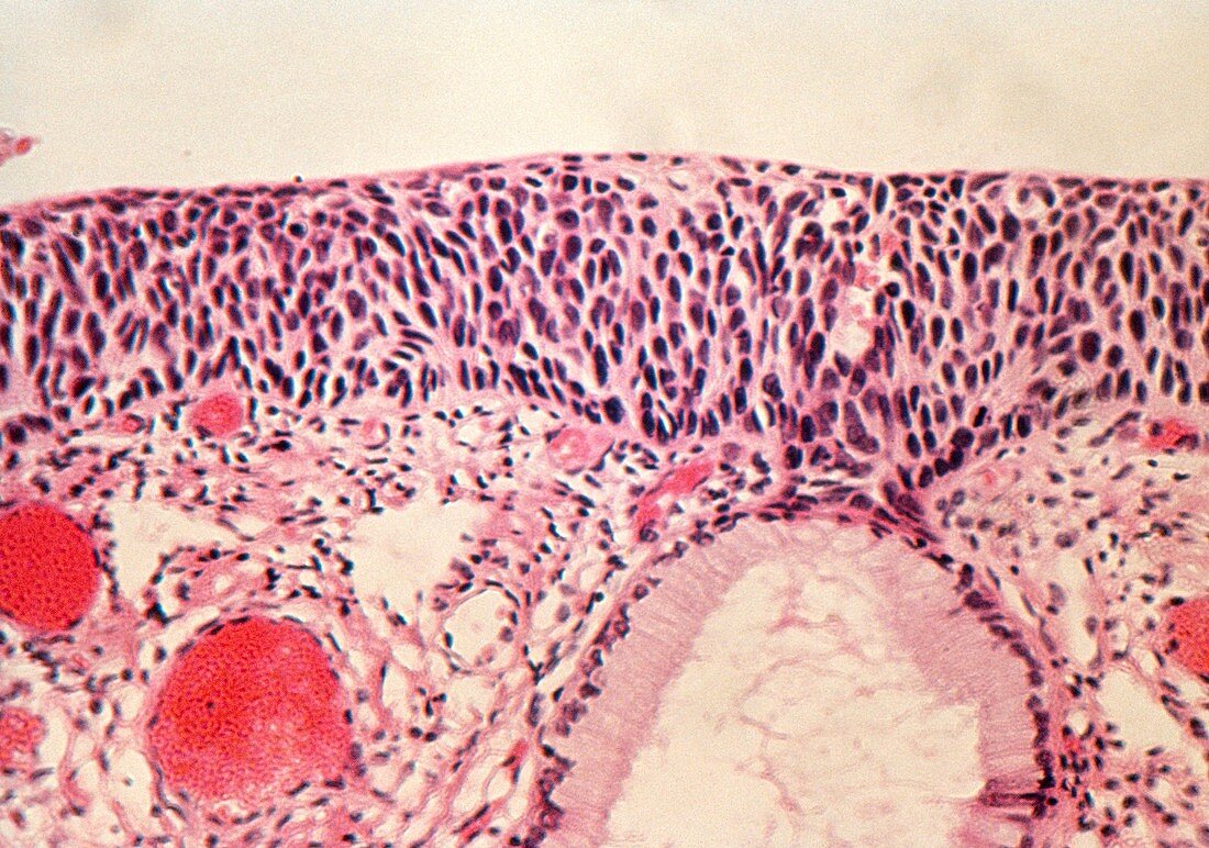
| Light micrograph of a biopsy section through the human cervix revealing carcinoma-in-situ (severe dysplasia,CIN 3). The cervix is lined by a stratified squamous epithelium. Normally,all cell division is confined to the basal epithelial layer (centre of image). As a cell matures & rises up the layers,its cytoplasm expands & nucleus shrinks in size - a normal superficial cell is flat,with a small nucleus & a large cytoplasm. In carcinoma-in-situ the gradual stratification in cell maturation seen in normal epithelium is absent: dysplastic (abnormal) cells extend throughout the entire epithelium. The basement membrane remains defined & intact | |
| Licence : | Droits gérés |
| Crédit: | Science Photo Library |
| Taille de l’image : | 5047 px × 3543 px |
| Model Release : | Non requis |
| Property Release : | Non requis |
| Restrictions : | - |
Prix pour cette image À partir de 45 €
Produit vendu
(Calendrier, Carte postale, Carte de vœux, Impression sur textile, Packaging etc)
À partir de 45 €
Usage commercial
(Affichage, Annonce presse, Annonce TV, Carte, Digital - hors rés. sociaux, Digital - rés. sociaux etc)
À partir de 45 €
Éditorial
(Digital, Journal, Livre, Livre pratique, Magazine, Télévision etc)
À partir de 60 €
Usage non-commercial
(Digital - hors rés. sociaux, Digital - rés. sociaux etc)
À partir de 120 €