Extracted molar
Numéro d’image : 11859178
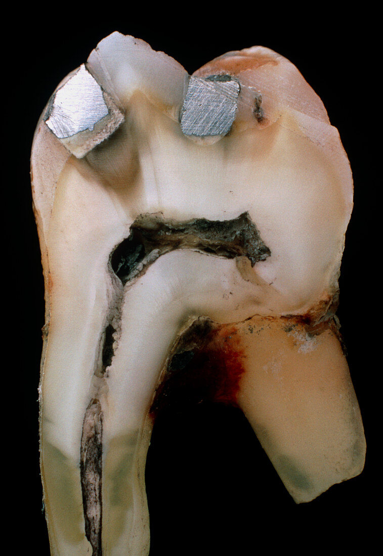
| Macrophotograph of an extracted molar (male,upper,number 7),sectioned with a glass cutting wheel. A root was broken during extraction. The molar shows two amalgam fillings,a powdered alloy consisting of silver,tin,copper & zinc mixed with mercury. The decayed area is removed & the cavity lined with a white filler (seen),which protects the base of the cavity from the effects of the filling itself. The open channel running up the root & across the tooth is the dental pulp containing blood vessels,nerves & lymphatics. A layer of enamel covers the crown,beneath which is the dentine (both seen here) running down the roots. Magnification:X 1.4 (at 35mm size) | |
| Licence : | Droits gérés |
| Crédit: | Science Photo Library / Burgess, Dr. Jeremy |
| Taille de l’image : | 3492 px × 5077 px |
| Model Release : | Non requis |
| Property Release : | Non requis |
| Restrictions : | - |
Prix pour cette image À partir de 45 €
Produit vendu
(Calendrier, Carte postale, Carte de vœux, Impression sur textile, Packaging etc)
À partir de 45 €
Usage commercial
(Affichage, Annonce presse, Annonce TV, Carte, Digital - hors rés. sociaux, Digital - rés. sociaux etc)
À partir de 45 €
Éditorial
(Digital, Journal, Livre, Livre pratique, Magazine, Télévision etc)
À partir de 60 €
Usage non-commercial
(Digital - hors rés. sociaux, Digital - rés. sociaux etc)
À partir de 120 €