Prosthetic knee
Numéro d’image : 11854113
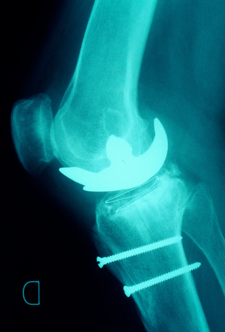
| Prosthetic knee. X-ray of a side view of a knee joint with an artificial or prosthetic joint (bright area). The head of the femur of the upper leg has been replaced with an implant. The tibia,the larger of the two bones in the lower leg,has been fitted with two screws,probably to pin the bone together during healing of a fracture. The kneecap or patella is also seen (far left). Knee-joint replacement may be carried out following an accident to the leg,or it may be necessary in patients severely affected by osteoarthritis or rheumatoid arthritis,which cause inflammation and erosion of the joints | |
| Licence : | Droits gérés |
| Crédit: | Science Photo Library / CNRI |
| Taille de l’image : | 3522 px × 5194 px |
| Model Release : | Non requis |
| Property Release : | Non requis |
| Restrictions : | - |
Prix pour cette image À partir de 45 €
Produit vendu
(Calendrier, Carte postale, Carte de vœux, Impression sur textile, Packaging etc)
À partir de 45 €
Usage commercial
(Affichage, Annonce presse, Annonce TV, Carte, Digital - hors rés. sociaux, Digital - rés. sociaux etc)
À partir de 45 €
Éditorial
(Digital, Journal, Livre, Livre pratique, Magazine, Télévision etc)
À partir de 60 €
Usage non-commercial
(Digital - hors rés. sociaux, Digital - rés. sociaux etc)
À partir de 120 €
Mots clés
- arthroplastie,
- articulation,
- broche,
- broches,
- chirurgical,
- coincé,
- dispositif prosthétique,
- dispositif prothétique,
- épinglé,
- fémur,
- fixée,
- genou,
- goupilles,
- implant artificiel,
- jambe,
- médecine,
- médical,
- médicale,
- membre,
- monochrome,
- n/b,
- noir et blanc,
- noir-et-blanc,
- os,
- pin,
- pins,
- prothèse,
- punaises,
- radiographie,
- rayons X,
- rechange,
- remplaçant,
- remplacement,
- réparation,
- réparer,
- soins de santé,
- tibia,
- tige,
- traitement,
- vis,
- vis métallique