Knee replacement,X-ray
Numéro d’image : 11854071
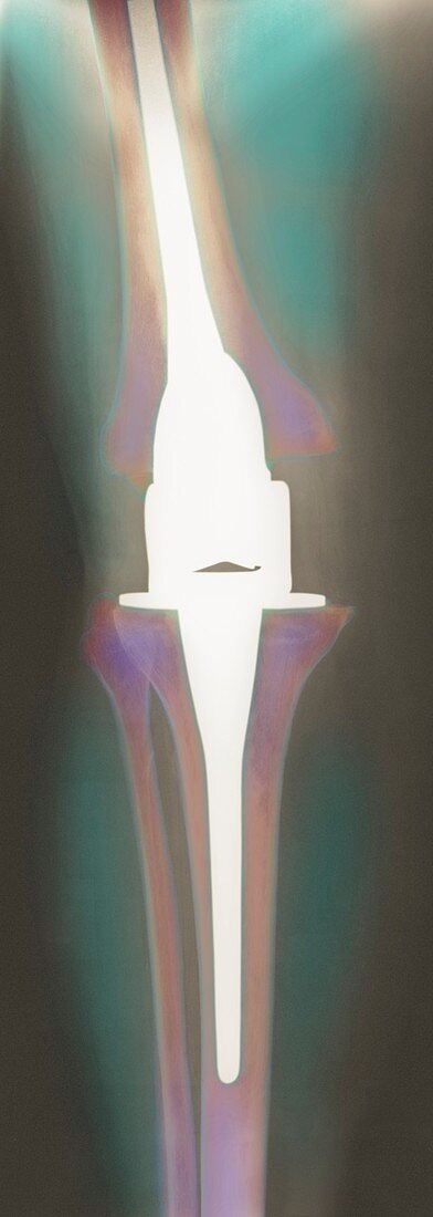
| Knee replacement. Coloured X-ray of a prosthetic knee implant (white),of a female patient's left leg (rear view). The implant is attached by pegs (at upper and lower centre) into the femur and tibia bones (brown/purple) respectively,with surgical cement. The flexible joint (centre) can hinge like the old joint. The patient's kneecap (patella,not seen) was not replaced. The implant replaced the old joint that had become damaged or lost cartilage,possibly due to an accident or osteoarthritis. Healthy cartilage reduces friction between the bones,and its progressive loss causes joint pain and immobility | |
| Licence : | Droits gérés |
| Crédit: | Science Photo Library |
| Taille de l’image : | 1879 px × 5276 px |
| Model Release : | Non requis |
| Property Release : | Non requis |
| Restrictions : | - |
Prix pour cette image À partir de 45 €
Produit vendu
(Calendrier, Carte postale, Carte de vœux, Impression sur textile, Packaging etc)
À partir de 45 €
Usage commercial
(Affichage, Annonce presse, Annonce TV, Carte, Digital - hors rés. sociaux, Digital - rés. sociaux etc)
À partir de 45 €
Éditorial
(Digital, Journal, Livre, Livre pratique, Magazine, Télévision etc)
À partir de 60 €
Usage non-commercial
(Digital - hors rés. sociaux, Digital - rés. sociaux etc)
À partir de 120 €
Mots clés
- arthrite,
- arthritis,
- arthrose,
- articulation,
- coloré,
- colorié,
- colorisé,
- cuisse,
- dispositif prosthétique,
- dispositif prothétique,
- féminin,
- féminine,
- femme,
- fémur,
- fibula,
- gauche,
- genou,
- implant,
- implant artificiel,
- jambe,
- jarret,
- médecine,
- médical,
- médicale,
- os,
- osteoarthritis,
- patient,
- patients,
- péroné,
- prothèse,
- radiographie,
- rayons X,
- rechange,
- remplaçant,
- remplacé,
- remplacement,
- restant,
- soins de santé,
- tibia,
- traitement,
- vue arrière