Brain surgeon uses MRI scanner during surgery
Numéro d’image : 11853260
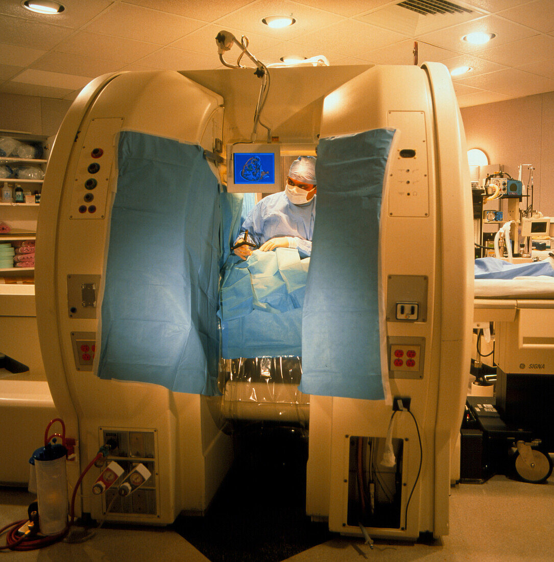
| 3-D virtual reality assisted surgery. Brain surgeon using an adapted MRI (magnetic resonance imaging) scanner to perform surgery to remove a tumour. He is aided by 3-D virtual reality (VR). On the monitor (at upper centre) is a 3-D VR model of the patient's brain. It shows internal structures of the brain in relation to the tumour (green). The image was computer generated from several MRI slice images through the brain. During surgery new MRI scans are taken so that the VR model can be adjusted to locate the precise place of the tumour. The tumour can thus be removed with the minimum of invasive surgery. Surgery at the Brigham and Women's Hospital,USA | |
| Licence : | Droits gérés |
| Crédit: | Science Photo Library / Ogden, Sam |
| Taille de l’image : | 2480 px × 2514 px |
| Model Release : | Le droit n'est pas encore disponible. Merci de nous contacter avant utilisation. |
| Property Release : | Non requis |
| Restrictions : |
|
Prix pour cette image À partir de 45 €
Produit vendu
(Calendrier, Carte postale, Carte de vœux, Impression sur textile, Packaging etc)
À partir de 45 €
Usage commercial
(Affichage, Annonce presse, Annonce TV, Carte, Digital - hors rés. sociaux, Digital - rés. sociaux etc)
À partir de 45 €
Éditorial
(Digital, Journal, Livre, Livre pratique, Magazine, Télévision etc)
À partir de 60 €
Usage non-commercial
(Digital - hors rés. sociaux, Digital - rés. sociaux etc)
À partir de 120 €
Mots clés
- cerveau,
- chirurgie du cerveau,
- I.R.M.,
- Imagerie par Résonance Magnétique,
- imagerie par résonnance magnétique,
- IRM,
- médecine,
- médical,
- médicale,
- neuro-imagerie,
- neurochirurgie,
- neuroimagerie,
- opération chirurgicale,
- R.V.,
- réalité virtuelle,
- RV,
- soins de santé,
- traitement chirurgical,
- tumeurs cérébrales