Doctor monitoring patient undergoing CT scan
Numéro d’image : 11848875
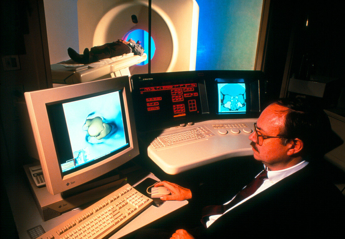
| CT scanning. Doctor overseeing the computed tomography (CT) scanning of a patient (upper left) from a control room. CT scanning uses X-rays to take slice images through the body (as seen on the monitor at upper right). Several slice images can be combined to create a three-dimensional (3-D) image,such as the one on the monitor at centre left,which shows an atheroma plaque blocking an artery. These 3-D views can be used to study the interior structures of the body without using invasive techniques. CT scanning is useful for detecting tumours in organs and can also be used to study smaller areas in detail,such as the insides of arteries | |
| Licence : | Droits gérés |
| Crédit: | Science Photo Library / Goetgheluck, Pascal |
| Taille de l’image : | 3637 px × 2516 px |
| Model Release : | Le droit n'est pas encore disponible. Merci de nous contacter avant utilisation. |
| Property Release : | Non requis |
| Restrictions : | - |
Prix pour cette image À partir de 45 €
Produit vendu
(Calendrier, Carte postale, Carte de vœux, Impression sur textile, Packaging etc)
À partir de 45 €
Usage commercial
(Affichage, Annonce presse, Annonce TV, Carte, Digital - hors rés. sociaux, Digital - rés. sociaux etc)
À partir de 45 €
Éditorial
(Digital, Journal, Livre, Livre pratique, Magazine, Télévision etc)
À partir de 60 €
Usage non-commercial
(Digital - hors rés. sociaux, Digital - rés. sociaux etc)
À partir de 120 €
Mots clés
- balayage CT 3D,
- CT scan,
- diagnostic,
- diagnostics,
- diagnostique,
- diagnostiques,
- équipement,
- matériel,
- médecin,
- médecine,
- médical,
- médicale,
- patient,
- patient dans,
- patients,
- radiographie,
- rayons X,
- salle de commandement,
- salle de controle,
- scan ct,
- scanner,
- scanner CT,
- soins de santé,
- T.D.M.,
- TDM,
- technique,
- tomodensitométrie,
- tomodensitométrie 3D,
- tomographie par ordinateur