Radiographer examining a series of CT scans
Numéro d’image : 11848855
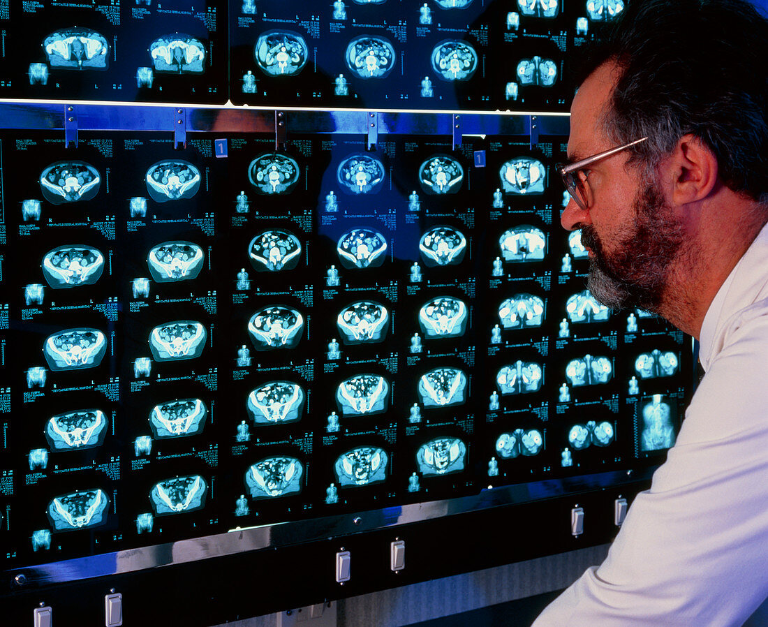
| MODEL RELEASED. Examining CT scans. Radiographer examines a series of computed tomography (CT) scans on a lightbox. These scans show successive "slices" through the pelvis of a patient,with bone coloured white. CT images are obtained from a series of X-ray beams passed through the patient. The scanner is known as a tomograph (from a Greek word meaning slice). CT scans display good contrast between soft tissues in the body,differentiating between bone,fat,muscle and other tissues. CT scans are useful in locating tumours,blood clots,and diagnosing diseases | |
| Licence : | Droits gérés |
| Crédit: | Science Photo Library / MAIN X-RAY, NEWCASTLE GENERAL HOSPITAL / SIMON FRASER |
| Taille de l’image : | 4672 px × 3815 px |
| Model Release : | Disponible |
| Property Release : | Non requis |
| Restrictions : | - |
Prix pour cette image À partir de 45 €
Produit vendu
(Calendrier, Carte postale, Carte de vœux, Impression sur textile, Packaging etc)
À partir de 45 €
Usage commercial
(Affichage, Annonce presse, Annonce TV, Carte, Digital - hors rés. sociaux, Digital - rés. sociaux etc)
À partir de 45 €
Éditorial
(Digital, Journal, Livre, Livre pratique, Magazine, Télévision etc)
À partir de 60 €
Usage non-commercial
(Digital - hors rés. sociaux, Digital - rés. sociaux etc)
À partir de 120 €