Doppler ultrasound scan
Numéro d’image : 11848758
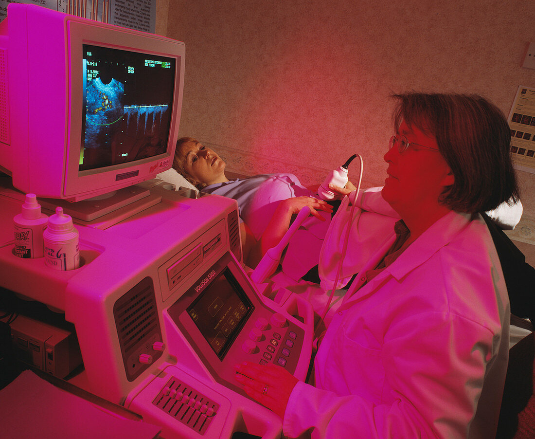
| MODEL RELEASED. Doppler ultrasound scan. Patient having a doppler ultrasound scan taken of her iliac artery during an investigation into blood flow. Ultrasound uses sound waves to build up an internal image of the body. A transducer held on the woman's abdomen emits high-frequency sound waves and picks up their reflections off body structures. This allows a 2-D or 3-D image of the structures to be created. Doppler ultrasound measures blood flow using the doppler effect. Sound waves reflected by moving objects have their frequencies shifted by an amount related to the speed of movement. The bright patches on the screen show the fastest blood flow | |
| Licence : | Droits gérés |
| Crédit: | Science Photo Library / McLean, John |
| Taille de l’image : | 4185 px × 3432 px |
| Model Release : | Disponible |
| Property Release : | Non requis |
| Restrictions : | - |
Prix pour cette image À partir de 45 €
Produit vendu
(Calendrier, Carte postale, Carte de vœux, Impression sur textile, Packaging etc)
À partir de 45 €
Usage commercial
(Affichage, Annonce presse, Annonce TV, Carte, Digital - hors rés. sociaux, Digital - rés. sociaux etc)
À partir de 45 €
Éditorial
(Digital, Journal, Livre, Livre pratique, Magazine, Télévision etc)
À partir de 60 €
Usage non-commercial
(Digital - hors rés. sociaux, Digital - rés. sociaux etc)
À partir de 120 €
Mots clés
- artère iliaque,
- balayage ultrason,
- capteur,
- diagnostic,
- diagnostics,
- diagnostique,
- diagnostiques,
- doppler,
- écoulement,
- écran,
- enregistrement,
- enregistrer,
- équipement,
- féminin,
- féminine,
- femme,
- femmes,
- gynécologie,
- gynécologue,
- matériel,
- médecin,
- médecine,
- médical,
- médicale,
- moniteur,
- ordinateur,
- scanner,
- soins de santé,
- technique,
- transducteur,
- unité d'affichage,
- unité de visualisation,
- vaisseau sanguin