Woman undergoing an ultrasound breast scan
Numéro d’image : 11848728
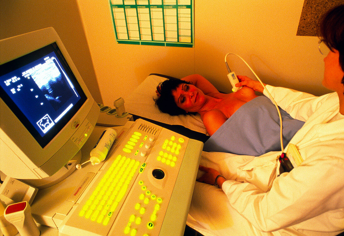
| Ultrasound breast scan. Female doctor carrying out an ultrasound breast scan. Ultrasound scans are produced using high-frequency sound waves from a hand-held transducer. The waves reflect back to the transducer off internal body structures and are formed into the real-time image seen on the monitor at far left. Ultrasound breast scans are used to distinguish between solid tumours and fluid-filled cysts. They are normally carried out once a mammogram (breast X-ray) has revealed an abnormality. With a tumour,a biopsy (tissue sampling) must be carried out to see whether it is cancerous. Photographed at the European Institute of Oncology in Milan,Italy | |
| Licence : | Droits gérés |
| Crédit: | Science Photo Library / Fermariello, Mauro |
| Taille de l’image : | 3617 px × 2480 px |
| Model Release : | Le droit n'est pas encore disponible. Merci de nous contacter avant utilisation. |
| Property Release : | Non requis |
| Restrictions : | - |
Prix pour cette image À partir de 45 €
Produit vendu
(Calendrier, Carte postale, Carte de vœux, Impression sur textile, Packaging etc)
À partir de 45 €
Usage commercial
(Affichage, Annonce presse, Annonce TV, Carte, Digital - hors rés. sociaux, Digital - rés. sociaux etc)
À partir de 45 €
Éditorial
(Digital, Journal, Livre, Livre pratique, Magazine, Télévision etc)
À partir de 60 €
Usage non-commercial
(Digital - hors rés. sociaux, Digital - rés. sociaux etc)
À partir de 120 €
Mots clés
- balayage à ultrasons,
- balayage ultrason,
- capteur,
- diagnostic,
- diagnostics,
- diagnostique,
- diagnostiques,
- échogramme,
- échographie,
- équipement,
- examen des seins,
- femme médecin,
- matériel,
- médecin,
- médecine,
- médical,
- médicale,
- moniteur,
- poitrine,
- projection,
- scanner,
- sein,
- soins de santé,
- technique,
- transducteur,
- ultrason