Abdominal ultrasound examination
Numéro d’image : 11848679
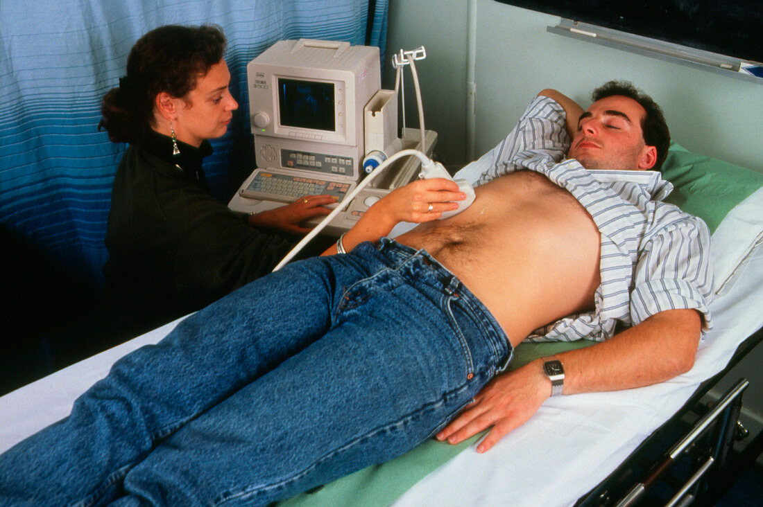
| Young man receiving an abdominal ultrasound scan. Ultrasonography is used to investigate a number of abdominal organs,notably the kidneys,urinary bladder,liver and pancreas. Here,an image of the patient's abdominal anatomy appears on the screen as the operator holds the scanner's transducer against the skin. The transducer emits high- frequency sound waves in short pulses & acts as a receiver for returning echoes,which occur when the ultrasound pulse encounters the interface between two tissue types. The technique is useful in distinguishing between renal solid tumours & fluid-filled cysts | |
| Licence : | Droits gérés |
| Crédit: | Science Photo Library / St. Bartholomew's Hospital |
| Taille de l’image : | 3526 px × 2346 px |
| Model Release : | Le droit n'est pas encore disponible. Merci de nous contacter avant utilisation. |
| Property Release : | Non requis |
| Restrictions : | - |
Prix pour cette image À partir de 45 €
Produit vendu
(Calendrier, Carte postale, Carte de vœux, Impression sur textile, Packaging etc)
À partir de 45 €
Usage commercial
(Affichage, Annonce presse, Annonce TV, Carte, Digital - hors rés. sociaux, Digital - rés. sociaux etc)
À partir de 45 €
Éditorial
(Digital, Journal, Livre, Livre pratique, Magazine, Télévision etc)
À partir de 60 €
Usage non-commercial
(Digital - hors rés. sociaux, Digital - rés. sociaux etc)
À partir de 120 €