Coloured MRI of knee meniscus degeneration
Numéro d’image : 11844247
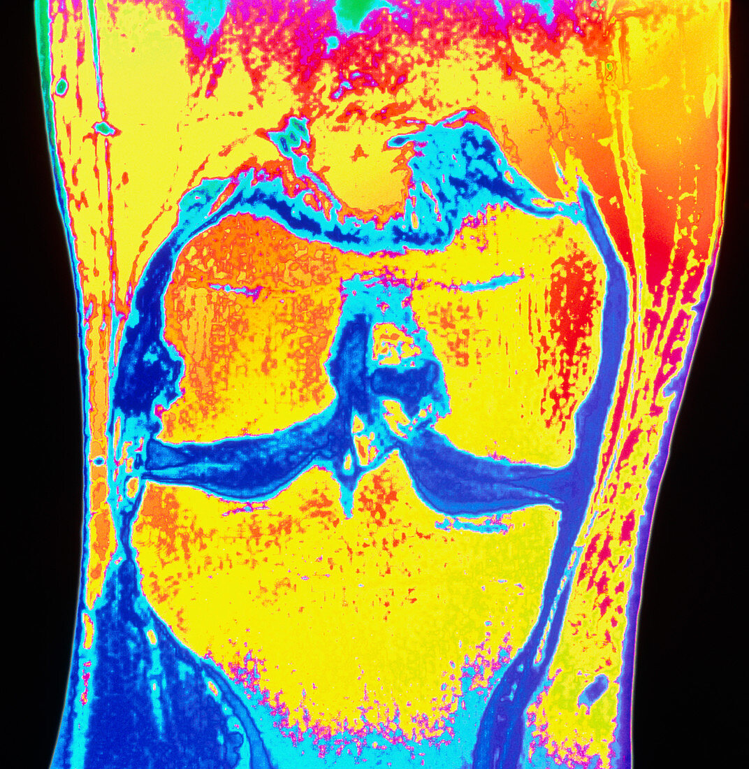
| Knee injury. Coloured Magnetic Resonance Image (MRI) scan of the knee joint of a 35 year old man,showing meniscus degeneration. The head of the femur (thigh bone,upper centre) articulates with the tibia (shin bone,lower centre) at a central dark area. This dark region consists of cartilage discs called menisci which provide a smooth,strong surface for movement. Due to compression of the medial meniscus cartilage,the tibia bone is damaged. This appears as a blue line in the bone near the joint surface (centre right). It is due to natural wear and tear of the joint,exacerbated by the patient's high activity in sport | |
| Licence : | Droits gérés |
| Crédit: | Science Photo Library / Fraser, Simon |
| Taille de l’image : | 4293 px × 4404 px |
| Model Release : | Non requis |
| Property Release : | Non requis |
| Restrictions : | - |
Prix pour cette image À partir de 45 €
Produit vendu
(Calendrier, Carte postale, Carte de vœux, Impression sur textile, Packaging etc)
À partir de 45 €
Usage commercial
(Affichage, Annonce presse, Annonce TV, Carte, Digital - hors rés. sociaux, Digital - rés. sociaux etc)
À partir de 45 €
Éditorial
(Digital, Journal, Livre, Livre pratique, Magazine, Télévision etc)
À partir de 60 €
Usage non-commercial
(Digital - hors rés. sociaux, Digital - rés. sociaux etc)
À partir de 120 €