PET scan (temporal) of carbon monoxide poisoning
Numéro d’image : 11844105
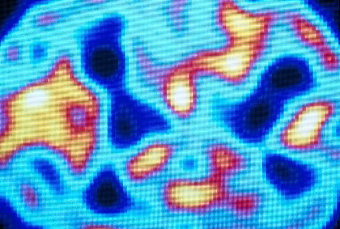
| Positron emission tomography (PET) scan of the brain (temporal lobe level) of a patient poisoned by carbon monoxide gas. This gas is common in car exhaust fumes. The colour-coded scan through this cerebral layer shows brain activity: from low (blue) to high (yellow). PET scanning relies on an injected radioactive tracer to reveal variations in metabolic activity in the brain. Normal brain activity produces a roughly symmetrical pattern in the yellow areas of left (upper) & right cerebral hemispheres. This scan shows reduced activity in one hemisphere. Carbon monoxide gas deprives brain tissues of oxygen,and may lead to permanent brain damage or even death | |
| Licence : | Droits gérés |
| Crédit: | Science Photo Library / Beddow, Tim |
| Taille de l’image : | 5150 px × 3471 px |
| Model Release : | Non requis |
| Property Release : | Non requis |
| Restrictions : | - |
Prix pour cette image À partir de 45 €
Produit vendu
(Calendrier, Carte postale, Carte de vœux, Impression sur textile, Packaging etc)
À partir de 45 €
Usage commercial
(Affichage, Annonce presse, Annonce TV, Carte, Digital - hors rés. sociaux, Digital - rés. sociaux etc)
À partir de 45 €
Éditorial
(Digital, Journal, Livre, Livre pratique, Magazine, Télévision etc)
À partir de 60 €
Usage non-commercial
(Digital - hors rés. sociaux, Digital - rés. sociaux etc)
À partir de 120 €