Tuberculosis
Numéro d’image : 11843258
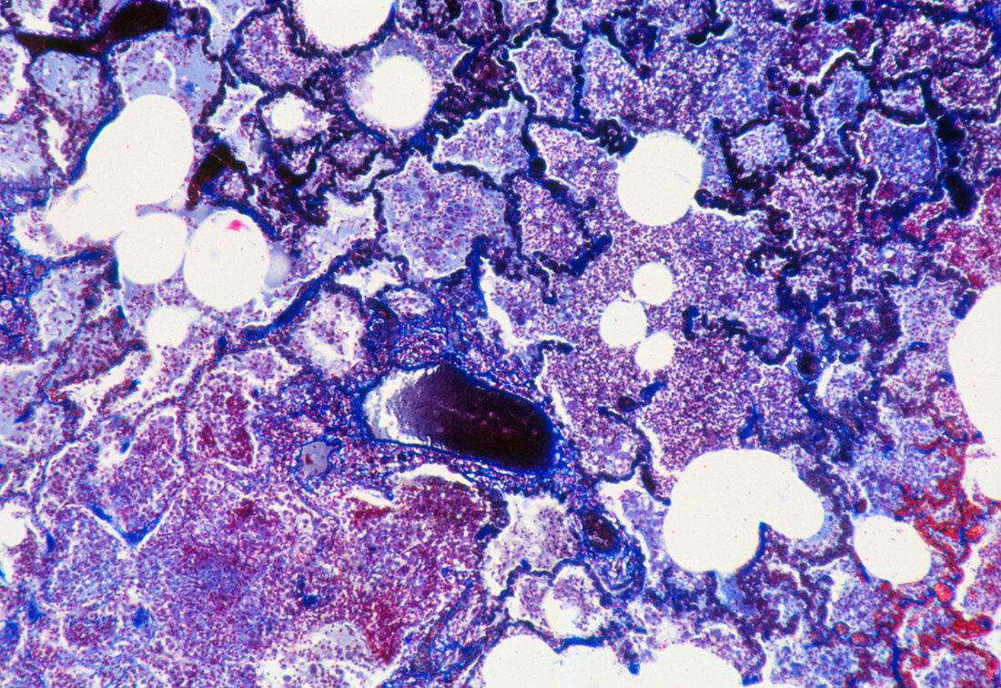
| Tuberculosis. Light micrograph of a human lung affected with tuberculosis. The air sac alveoli of the lung can be seen,filled with an inflammatory exudate. This exudate consists largely of macro- phages (white blood cells) and the infecting bacteria Mycobacterium tuberculosis. At lower right are numerous blood vessels (red); the walls of alveoli have disintegrated (at lower left). Tuberculosis is an infectious lung disease which once was a major killer disease worldwide. In the lungs,bacteria breathed in form inflammatory lesions known as "tubercles" which destroy lung tissue. Magnification: x20 at 35mm size | |
| Licence : | Droits gérés |
| Crédit: | Science Photo Library / Michler, Astrid & Hans-Frieder |
| Taille de l’image : | 3609 px × 2480 px |
| Model Release : | Non requis |
| Property Release : | Non requis |
| Restrictions : | - |
Prix pour cette image À partir de 45 €
Produit vendu
(Calendrier, Carte postale, Carte de vœux, Impression sur textile, Packaging etc)
À partir de 45 €
Usage commercial
(Affichage, Annonce presse, Annonce TV, Carte, Digital - hors rés. sociaux, Digital - rés. sociaux etc)
À partir de 45 €
Éditorial
(Digital, Journal, Livre, Livre pratique, Magazine, Télévision etc)
À partir de 60 €
Usage non-commercial
(Digital - hors rés. sociaux, Digital - rés. sociaux etc)
À partir de 120 €