Coloured MRI brain scan of Sturge-Weber syndrome
Numéro d’image : 11843056
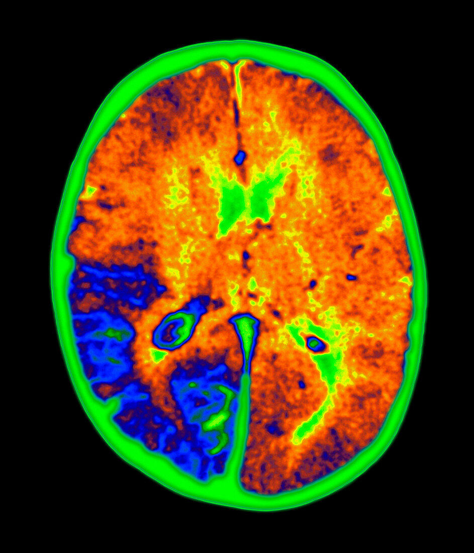
| Sturge-Weber syndrome. Coloured magnetic resonance imaging (MRI) scan of a brain (orange/blue) with Sturge-Weber syndrome. The brain is seen in horiz- ontal (axial) section,with the front of the brain at top. The brain's fluid-filled ventricles (upper centre & lower right) are green. Part of the brain has become calcified (blue,lower left). Other symptoms include angioma tumours in the choroid of the eye and the meninges membranes that cover the brain,as well as port-wine nevi on the face and glaucoma of the eye. People with this congenital disorder are mentally handicapped and may suffer from epileptic seizures. MRI uses radio waves and a powerful magnet to produce slice body images | |
| Licence : | Droits gérés |
| Crédit: | Science Photo Library / Kulyk, Mehau |
| Taille de l’image : | 3000 px × 3500 px |
| Model Release : | Non requis |
| Property Release : | Non requis |
| Restrictions : | - |
Prix pour cette image À partir de 45 €
Produit vendu
(Calendrier, Carte postale, Carte de vœux, Impression sur textile, Packaging etc)
À partir de 45 €
Usage commercial
(Affichage, Annonce presse, Annonce TV, Carte, Digital - hors rés. sociaux, Digital - rés. sociaux etc)
À partir de 45 €
Éditorial
(Digital, Journal, Livre, Livre pratique, Magazine, Télévision etc)
À partir de 60 €
Usage non-commercial
(Digital - hors rés. sociaux, Digital - rés. sociaux etc)
À partir de 120 €
Mots clés
- calcification,
- cerveau,
- coupé,
- coupe-circuit,
- découpé,
- découpes,
- désordre,
- détourages,
- détouré,
- difformité,
- disjoncteur,
- état,
- I.R.M.,
- imagerie par résonnance magnétique,
- IRM,
- maladie,
- médecine,
- médical,
- médicale,
- neuro-imagerie,
- neuroimagerie,
- retard,
- retardement,
- scanner du cerveau,
- silhouette,
- soins de santé,
- trouble