Coloured X-ray of pneumothorax treatment
Numéro d’image : 11841828
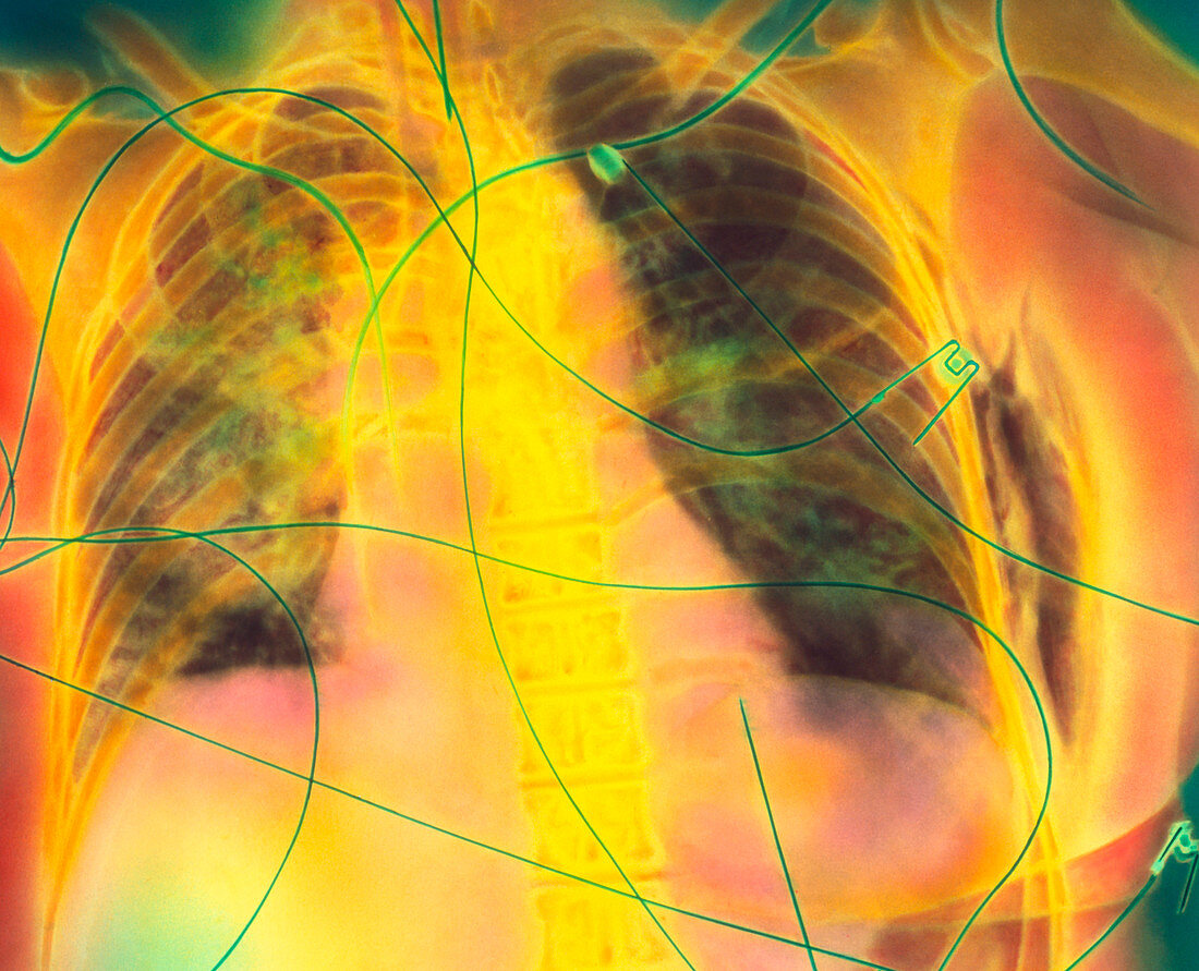
| Pneumothorax treatment. Coloured chest X-ray of a 51 year old woman during pneumothorax treatment,showing lung shadowing and surgical emphysema. Bones of the ribcage are seen. The darker lungs reveal extensive shadowing (lighter patches) but there is no longer evidence of pneumothorax. Pneumothorax occurs when air enters the pleural cavity around the lungs & the lungs may collapse. At right,surgical emphysema is seen outside the ribcage (dark area). ECG electrode leads attached to the chest are seen. An endotracheal tube is at top centre (green); central venous lines (upper left,top right) lead into major blood vessels | |
| Licence : | Droits gérés |
| Crédit: | Science Photo Library |
| Taille de l’image : | 3247 px × 2629 px |
| Model Release : | Non requis |
| Property Release : | Non requis |
| Restrictions : | - |
Prix pour cette image À partir de 45 €
Produit vendu
(Calendrier, Carte postale, Carte de vœux, Impression sur textile, Packaging etc)
À partir de 45 €
Usage commercial
(Affichage, Annonce presse, Annonce TV, Carte, Digital - hors rés. sociaux, Digital - rés. sociaux etc)
À partir de 45 €
Éditorial
(Digital, Journal, Livre, Livre pratique, Magazine, Télévision etc)
À partir de 60 €
Usage non-commercial
(Digital - hors rés. sociaux, Digital - rés. sociaux etc)
À partir de 120 €
Mots clés
- bronchopneumopathie chronique obstructive,
- buste,
- cage thoracique,
- chirurgical,
- désordre,
- ECG,
- électrocardiogramme,
- électrode,
- emphysema,
- emphysème,
- état,
- ligne veineuse centrale,
- maladie,
- médecine,
- médical,
- médicale,
- MPOC,
- pneumothorax,
- poumon,
- radiographie,
- rayons X,
- soins de santé,
- traitement,
- trouble