Coloured venogram of thrombosis in vein in the leg
Numéro d’image : 11840182
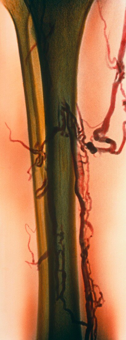
| Thrombosis. Coloured venogram (X-ray) showing deep vein thrombosis in the leg's popliteal vein (red,down right). The tibia (shin-bone,olive green) is beside the fibula (centre left). A large thrombus,or abnormal blood clot (dark red,centre right),is in the calf. There are also other smaller clots (dark red) causing irregular blood flow in branching veins of the leg. In thrombosis,a blood clot forms in an undamaged blood vessel,causing pain and tissue damage. Although thrombi often break up on their own,drugs can aid this process and surgery may be needed. A venogram is produced by injecting an X-ray opaque dye into a vein | |
| Licence : | Droits gérés |
| Crédit: | Science Photo Library |
| Taille de l’image : | 2585 px × 6946 px |
| Model Release : | Non requis |
| Property Release : | Non requis |
| Restrictions : | - |
Prix pour cette image À partir de 45 €
Produit vendu
(Calendrier, Carte postale, Carte de vœux, Impression sur textile, Packaging etc)
À partir de 45 €
Usage commercial
(Affichage, Annonce presse, Annonce TV, Carte, Digital - hors rés. sociaux, Digital - rés. sociaux etc)
À partir de 45 €
Éditorial
(Digital, Journal, Livre, Livre pratique, Magazine, Télévision etc)
À partir de 60 €
Usage non-commercial
(Digital - hors rés. sociaux, Digital - rés. sociaux etc)
À partir de 120 €
Mots clés
- cavographie,
- désordre,
- état,
- fibula,
- jambe,
- maladie,
- médecine,
- médical,
- médicale,
- péroné,
- phlébite profonde,
- soins de santé,
- thrombeuse veineuse profonde,
- thrombophlébite,
- thrombose,
- thrombose veineuse profonde,
- thrombosis,
- thrombus,
- tibia,
- trouble,
- vaisseau sanguin,
- vasculaire,
- veine,
- veine poplitée,
- veine profonde