Coloured X-ray of leg showing deep vein thrombosis
Numéro d’image : 11840154
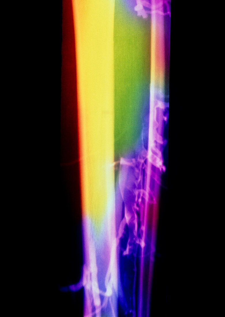
| Deep vein thrombosis. Coloured X-ray of a patient's lower leg showing deep vein thrombosis. The patient has been injected with an X-ray-opaque substance,making blood vessels show lighter. The large bone at left is the tibia; at right is the smaller fibula. A fresh clot (thrombus) has formed in the lower centre,where the blood vessels look concentrated. This has blocked a vein,preventing normal blood flow in the leg. Normal blood vessels reappear at the top. Deep vein thrombosis is blood clot formation in deep-lying veins. It is dangerous if the clot is large,as fragments may break off and block vital arteries elsewhere | |
| Licence : | Droits gérés |
| Crédit: | Science Photo Library |
| Taille de l’image : | 3480 px × 4909 px |
| Model Release : | Non requis |
| Property Release : | Non requis |
| Restrictions : | - |
Prix pour cette image À partir de 45 €
Produit vendu
(Calendrier, Carte postale, Carte de vœux, Impression sur textile, Packaging etc)
À partir de 45 €
Usage commercial
(Affichage, Annonce presse, Annonce TV, Carte, Digital - hors rés. sociaux, Digital - rés. sociaux etc)
À partir de 45 €
Éditorial
(Digital, Journal, Livre, Livre pratique, Magazine, Télévision etc)
À partir de 60 €
Usage non-commercial
(Digital - hors rés. sociaux, Digital - rés. sociaux etc)
À partir de 120 €